
Notes
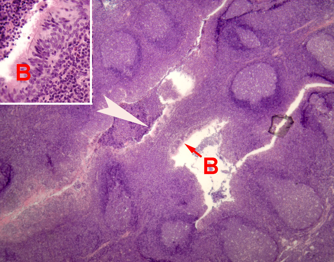
A tonsil consists of an epithelially-lined crypt (invaginated pocket) surrounded by dense clusters of lymph nodules, each with a germinal center where lymphocytes proliferate. The nodules are embedded in a mass of diffuse lymphoid tissue that consists of lymphocytes migrating to and from the germinal centers.
This tonsil of the pharynx is lined by pseudostratified columnar epithelium (B in image above). A dense infiltrate of lymphocytes occupies the mucosa. Lymphocytes may also be seen in the crypt lumen.
The mucosal-associated lymphoid tissue of the pharyngeal tonsils is similar to that of tonsils associated with the oral cavity (thumbnail links on right) as well as Peyer's patches and appendix. These structures, together with other more diffuse lymphoid tissue, constitute Mucosa-Associated Lymphoid Tissues, or MALT.
For more on MALT, for Mucosa-Associated Lymphoid Tissues), consult your histology text (e.g. pp. 134-5 in Stevens & Lowe).
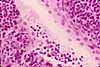
Related examples:
|
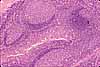 |
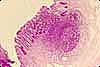 |
Comments and questions: dgking@siu.edu
SIUC / School
of Medicine / Anatomy / David
King
https://histology.siu.edu/crr/mq17.htm
Last updated: 26 May 2022 / dgk