Notes
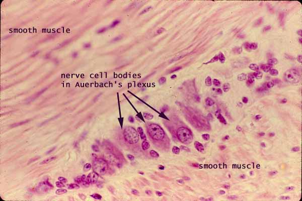
The muscularis externa consists of smooth muscle fibers arranged into two layers. The layer of circular muscle fibers (cut in longitudinal section) appears at the upper left, while the layer of longitudinal muscle fibers (cut in cross section) appears at lower right.
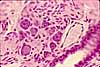
In between the two layers of muscle lies the myenteric nerve plexus, an interconnected network of parasympathetic ganglia which is also called "Auerbach's plexus" (after Leopold Auerbach, b. 1828). In routine histological specimens, the ganglia (small clusters of nerve cell bodies) are the only noticeable feature of Auerbach's plexus.
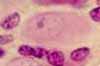
Nerve cell bodies are usually rather conspicuous. Each cell body can be quite large (up to ~50µm), with relatively basophilic cytoplasm, with a large round euchromatic nucleus, and with a single prominent nucleolus (one cell body is emphasized in the small image at left).
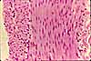
Associated with peripheral ganglia are numerous small satellite cells and Schwann cells (several of these are emphasized in the small image at right), as well as fibroblasts of the surrounding connective tissue.
Related examples:
 |
 |
 |
 |
||
Comments and questions: dgking@siu.edu
SIUC / School
of Medicine / Anatomy / David
King
https://histology.siu.edu/erg/GI010b.htm
Last updated: 14 May 2022 / dgk