
Notes
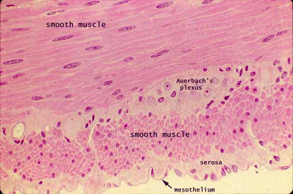
The muscularis externa of the small intestine consists of smooth muscle fibers arranged into two layers.
In the image above, the circular muscle fibers (in the upper half of the image) are cut longitudinally, while the longitudinal muscle fibers are cut in cross section (transversely). (More on appearance of smooth muscle.)
Between these two muscle layers is a network of unmyelinated nerve fibers and parasympathetic ganglia called Auerbach's plexus (or myenteric plexus).
The nerve cells in the micrograph above are not well-stained, although typical "owl-eye" or "fried-egg" nuclei (i.e., euchromatic with a prominent nucleolus) are visible on either end of the word "plexus".
The serosa consists of connective tissue with a delicate covering of mesothelium (simple squamous epithelium derived from mesoderm).
Related examples:
 |
 |
 |
|
||
Comments and questions: dgking@siu.edu
SIUC / School
of Medicine / Anatomy / David
King
https://histology.siu.edu/erg/GI011b.htm
Last updated: 17 May 2022 / dgk