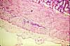
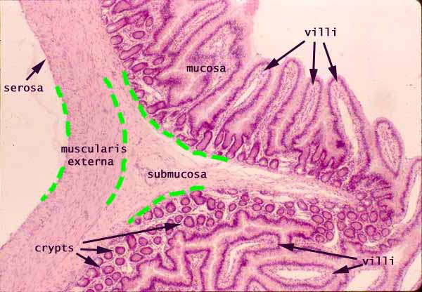
Jejunum, basic layers with plica (cross section)
Notes
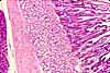
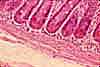
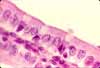
The mucosa of the jejunum is typical of the small intestine, with villi and crypts.
In this specimen, an intestinal plica with its submucosal core is conspicuous across the center of the image.
This specimen also clearly displays how crypts and villi can take on different appearances depending on plane of section.
Notice in this micrograph how the changing orientation of the mucosa with respect to the plane of section reveals the three-dimensional shape of villi and crypts.
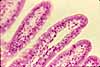
In the upper half of the image (on "top" of the plica), the mucosa is cut in cross section so that villi appear as fingers and crypts appear as invaginations. This is the "standard textbook view" of intestinal mucosa.
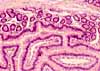
In the lower half of the image ("below" the plica), the mucosa is cut obliquely so that villi appear as floating "islands" of tissue (with a core of lamina propria) while crypts appear as small "donuts" embedded within the mucosa (surrounded by lamina propria.
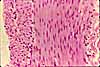
Muscularis externa of the jejunum has the standard inner circular and outer longitudinal layers of smooth muscle.
The serosa is too thin to appear on this low-magnification image, although its location is indicated.
Related examples:
Comments and questions: dgking@siu.edu
SIUC / School
of Medicine / Anatomy / David
King
https://histology.siu.edu/erg/GI031b.htm
Last updated: 14 May 2022 / dgk