Tunics and mediastinum
 Each
testis is suspended in an outpouching of the peritoneal cavity. This
cavity, like the rest of the peritoneum, is lined by a serosa
consisting of mesothelial cells supported
by fibrous connective tissue. Around the testis, the parietal peritoneum
is named tunica vaginalis while the visceral peritoneum is named tunica
albuginea. Both of these tunics consist of fibrous connective tissue
with a thin surface of mesothelium.
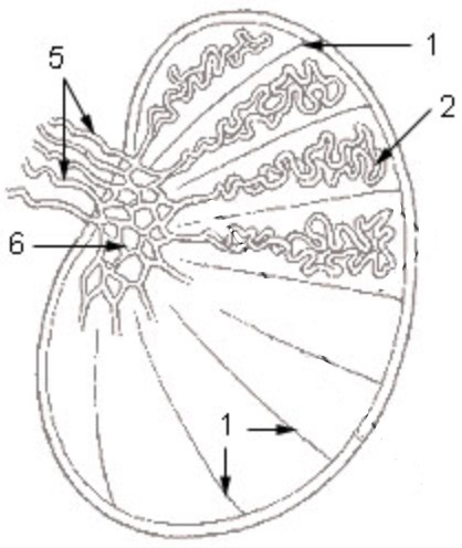
The thickened posterior portion of the tunica albuginea,
called the mediastinum, receives the blood vessels, lymphatics, nerves,
and ducts which serve the testis. Fibrous septa extend from the
mediastinum into the body of the testis.
Seminiferous tubules
 The
bulk of each testis consists of seminiferous tubules embedded in relatively
sparse interstitial tissue. Sperm
cells are produced by the tubules, while hormones are produced by endocrine
cells (Leydig cells) within the interstitium.
Tubules are surrounded by a thin layer of contractile myoid
cells.
Unlike the tubules in a typical exocrine gland, each seminiferous
tubule forms a tightly coiled loop, nearly a meter in length, which opens
at both ends into the rete testis.
Although such organization into loops is unique, the resulting
appearance in tissue section is typical for a tubular gland -- lots of round
or oval slices across tubules, with occasional tangential sections of the
epithelium.
A few hundred tubules comprise one testis. Thin connective
tissue septa, arising in the mediastinum, separate tubules into lobules.
Infertility due to atrophy of the testicular tubules
can result from a number of causes, ranging from undescended testes (cryptorchidism)
to inflammation (orchitis) to malnutrition or hormonal imbalance.
For images, see WebPath
and WebPath
(or Milikowski & Berman's Color Atlas of Basic Histopathology).
Sertoli cells
 Seminiferous tubules are lined by a complex epithelium which is most easily understood
as consisting of two very different cell populations, Sertoli cells
and germ cells.
Sertoli cells (the name commemorates Enrico Sertoli,
b. 1842) are support cells. These cells form what is essentially
a simple columnar epithelium.
Each Sertoli cell rests on the basement membrane and extends to the lumen.
The Sertoli cells create the environment in which germ cells carry out the fundamental
reproductive function of gamete production.
Intercellular junctions near the base of adjacent Sertoli cells create the
blood-testis barrier.
Spermatogonia lie on the "blood" side of this barrier. As these cells begin mitosis, spermatocytes
must cross the barrier; thus this barrier undergoes constant remodelling during spermatogenesis.
The simple columnar nature of the Sertoli epithelium is
most evident prior to puberty, before spermatogonia begin meiotic activity.
 In an adult testis, Sertoli cell nuclei are often inconspicuous among
the much more numerous nuclei of germ cells in various stages of meiosis and
maturation. Nevertheless, the nuclei of Sertoli cell can be readily
recognized as those typical of columnar epithelium -- oval, euchromatic nuclei,
usually with prominent nucleoli.
The cytoplasm of Sertoli cells assumes an elaborate shape,
enveloping germ cells at various stages in meiosis.
Primary spermatocytes, produced by mitosis
from spermatogonia, move away from the base of the
epithelium and are sealed off from the basal surface by tight junctions between
Sertoli cells. This has the effect of separating haploid germ
cells, which are antigenically foreign, from circulating antibodies. As
meiosis proceeds toward the epithelial surface, the germ cells
remain nestled in pockets in the Sertoli cell cytoplasm. Specific Sertoli
cell functions include nutritional support, phagocytosis of residual bodies
(shed cytoplasm) from spermatids, and secretion.
Germ cells
and Sperm cell formation
 Male
germ cells comprise a unique cell population which continually
produces new male gametes, or spermatozoa, in the process called
spermatogenesis. Germ cells at all stages of meiosis are
found embedded within the epithelium of the seminiferous
tubules.
|
Animation from Blue
Histology, Copyright
Lutz Slomianka 1998-2004
(The image should be animated,
if you watch patiently.)
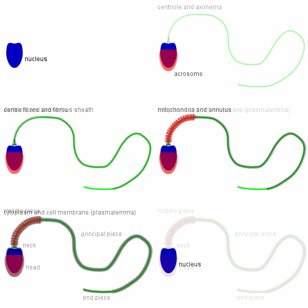
 The
mature human spermatozoon is about 60 Ám long and actively motile.
It is divided into head, neck and tail. The
mature human spermatozoon is about 60 Ám long and actively motile.
It is divided into head, neck and tail.
The head (flattened, about 5 Ám long and 3 Ám wide) chiefly
consists of the nucleus (greatly condensed chromatin!). The anterior
2/3 of the nucleus is covered by the acrosome, which contains
enzymes important in the process of fertilisation. The posterior
part of the nuclear membrane forms the so-called basal plate.
The neck is short (about 1 Ám) and attached to the basal
plate. A transversely oriented centriole is located immediately
behind the basal plate. The neck also contains nine segmented
columns of fibrous material, which continue as the outer dense
fibres into the tail.
The tail is further divided into a middle piece,
a principal piece and an end piece. The axonema
(the generic name for the arrangement of microtubules in all cilia)
begins in the middle piece. It is surrounded by nine outer dense
fibres, which are not found in other cilia. In the middle piece
(about 5 Ám long), the axonema and dense fibres are surrounded
by a sheath of mitochondria. The middle piece is terminated by
a dense ring, the annulus. The principal piece is about 45 Ám
long. It contains a fibrous sheath, which consists of dorsal and
ventral longitudinal columns interconnected by regularly spaced
circumferential hoops. The fibrous sheath and the dense fibres
do not extend to the tip of the tail. Along the last part (5 Ám)
of the tail, called the end piece, the axonema is only surrounded
by a small amount of cytoplasm and the plasma membrane.
|
For images of tumors involving
germ cells (seminomas and carcinomas), see WebPath
and WebPath
or Milikowski & Berman's Color Atlas of Basic Histopathology,
pp. 428-435.
-
Spermatogonia ...
-
are the stem cells of the germ cell population.
-
divide mitotically to produce primary spermatocytes
as well as more spermatogonia.
-
are found at the base of the tubular epithelium.
-
have relatively large round nuclei and lie
adjacent to the basement membrane of the tubular epithelium.
-
Primary spermatocytes ...
-
are cells in the first stage in meiosis, during
which DNA replicates twice.
- divide to produce secondary spermatocytes.
- have relatively large round nuclei.
- are found at mid-levels within the tubular epithelium.
-
Secondary spermatocytes ...
-
divide one more time, without further DNA replication,
to produce spermatids.
- have relatively large round nuclei (like primary spermatocytes).
- are found at mid-levels within the tubular epithelium (like primary
spermatocytes).
-
Spermatids ...
-
are the product of the final meiotic division.
- are found near the lumen of the tubule.
- have small round nuclei.
-
undergo an elaborate process of maturation (called
spermiogenesis) to become spermatozoa.
-
Spermatozoa ...
-
are highly specialized, motile cells, each with
a single large flagellum.
-
form by maturation (i.e., without further cell
division) from spermatids.
-
have very small, highly condensed, oval to conical
nuclei. (The shape of entire sperm cells, with long flagella,
is not evident in tissue sections.)
- are found near the lumen of the tubule.
- (See your histology textbook for more detailed information
on spermatozoa, including important features such as the acrosome, the
middle piece, and the axoneme, and the process of spermiogenesis.)
With fine histological material and exquisite attention
to nuclear detail, one may not only distinguish among spermatogonia, primary
spermatocytes, secondary spermatocytes, spermatids, and spermatozoa, but
also distinguish various stages of cell division within each cell type.
Historical note: Because
reproduction was once one of the grandest mysteries of life, the process
of gamete production has long received intense attention from biologists.
One major discovery during the late 1800s
was the marvelous dance of chromosomes during
meiosis. The profound significance of this process became apparent
with the rediscovery of Mendel's laws, when biologists realized that
the segregation and independent assortment of "genetic factors"
(what we now call genes) paralleled the behavior chromosomes
during meiosis. Chromosomes were the visible, physical agents
of inheritance!
With such a history, it should come as no surprise
that science has accumulated a vast amount of detailed information describing
both spermatogenesis and spermiogenesis. Growth of knowledge continues
apace, now motivated less by a sense of profound mystery than by an
expectation that a sufficiently detailed understanding the process might
yield better methods of birth control.
 Testosterone-secreting Leydig cells (named after Franz
von Leydig, b. 1821), also called "interstitial cells," occur in clusters within the interstitial tissue
(stroma) of the testis.
Leydig cells may be recognized not only by their location
within the testicular interstitium but also by their round nuclei
and extensive acidophilic cytoplasm.
Leydig cells have an appearance typical of steroid-secreting
cells. With electron microscopy they can be seen to contain
abundant smooth endoplasmic reticulum and mitochodria with tubular
cristae. Leydig cells may contain small eosinophilic cytoplasmic
inclusions called Reinke's crystaloids. With age, Leydig
cells may accumulate lipofuscin (brown "wear-and-tear" pigment).
 Each
testis is suspended in an outpouching of the peritoneal cavity. This
cavity, like the rest of the peritoneum, is lined by a serosa
consisting of mesothelial cells supported
by fibrous connective tissue. Around the testis, the parietal peritoneum
is named tunica vaginalis while the visceral peritoneum is named tunica
albuginea. Both of these tunics consist of fibrous connective tissue
with a thin surface of mesothelium.
 The
bulk of each testis consists of seminiferous tubules embedded in relatively
sparse interstitial tissue. Sperm
cells are produced by the tubules, while hormones are produced by endocrine
cells (Leydig cells) within the interstitium.
Tubules are surrounded by a thin layer of contractile myoid
cells.
 Seminiferous tubules are lined by a complex epithelium which is most easily understood
as consisting of two very different cell populations, Sertoli cells
and germ cells.
 In an adult testis, Sertoli cell nuclei are often inconspicuous among
the much more numerous nuclei of germ cells in various stages of meiosis and
maturation. Nevertheless, the nuclei of Sertoli cell can be readily
recognized as those typical of columnar epithelium -- oval, euchromatic nuclei,
usually with prominent nucleoli.


