
Notes
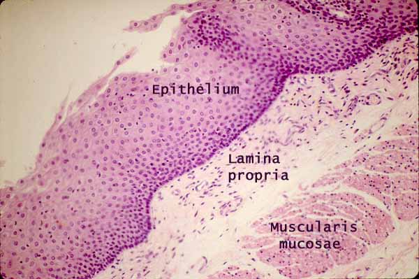
In the mucosa of the esophagus --
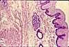
Esophageal epithelium is non-keratinized stratified squamous.
To recognize this epithelium as non-keratinized, compare with epidermis of skin while noting here the absence of a stratum granulosum, and note that cells on the lumenal surface of this epithelium appear similar to those deeper in the epithelium, with nuclei clearly present.
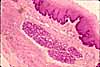
Esophageal lamina propria is less cellular (fewer lymphocytes) than that in the stomach and intestine, presumably because the protective stratified squamous epithelium is more effective at keeping out foreign antigens. Nevertheless, lymph nodules may occur (see thumbnail at right).
Esophageal muscularis mucosa is noticably thicker than that in the stomach and intestine, but includes only longitudinal muscle fibers.
When the esophagus is cut in cross section (across its long axis, as in the micrograph above), all of the smooth muscle fibers in the muscularis mucosae will also be cut in cross section. (Hence, all the smooth muscle nuclei in the above micrograph appear small and round, rather than elongated.)
Because the longitudinal fibers occur in bundles, a longitudinal section passing between bundles may not include any evidence of muscularis mucosae.
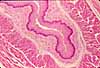
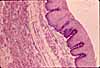
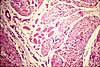
More esophagus examples:
 |
 |
 |
 |
 |
|
Comments and questions: dgking@siu.edu
SIUC / School
of Medicine / Anatomy / David
King
https://histology.siu.edu/erg/Gi006b.htm
Last updated: 27 May 2022 / dgk