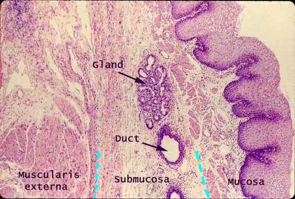

Esophagus, basic layers (cross section)

Notes
In the mucosa of the esophagus --
Esophageal epithelium is non-keratinized stratified squamous. Note that the basal surface of the epithelium is deeply indented by connective tissue papillae.
In oblique section through the epithelium, the connective tissue papillae can look like "islands," apparently surrounded by epithelium. Beginning students frequently mistake such an appearance for glands.
Esophageal lamina propria is less cellular (fewer lymphocytes) than lamina propria in the stomach and intestine, presumably because the protective stratified squamous epithelium is more effective at keeping out foreign antigens. Nevertheless, lymph nodules may occur in esophageal mucosa (thumbnail at right).
Esophageal muscularis mucosa is noticably thicker than that in the stomach and intestine, and includes only longitudinal muscle fibers.
Because the longitudinal fibers occur in bundles, a longitudinal section passing between bundles may not include any evidence of muscularis mucosae.
Esophageal submucosa includes scattered esophageal glands, which may or may occur in any given specimen.
Muscularis externa of the esophagus consists of the standard inner circular and outer longitudinal layers of smooth muscle, with Auerbach's plexus in between. Only the circular muscle is included in this micrograph (at lower left corner, interrupted by connective tissue at upper left).
More esophagus examples:
 |
 |
 |
 |
 |
Comments and questions: dgking@siu.edu
SIUC / School
of Medicine / Anatomy / David
King
https://histology.siu.edu/erg/GI060b.htm
Last updated: 9 May 2022 / dgk