
Notes
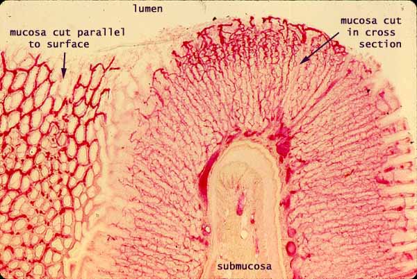
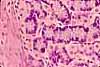
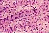
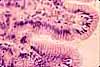
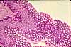
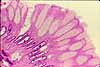
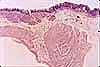
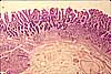
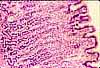
This image shows an unstained section of the stomach, after red dye had been injected into the vascular system. The mucosal architecture, with gastric pits and closely-packed tubular glands, is evident simply through the distribution of blood vessels.
Note the extensive capillary bed thoughout the mucosa. Capillaries are located in lamina propria between the gastric glands. (These capillaries are not always evident in routine sections.)
Also note that larger vessels are located at the base of the mucosa. (Much larger vessels, not included in this view, are located still deeper, in the submucosa. The latter are served by still larger vessels outside the muscularis externa.)
At the left, where the mucosa has been cut parallel to its surface, capillaries can be seen encircling gastric pits.
Related examples:
 |
 |
 |
 |
 |
 |
 |
|
 |
 |
 |
 |
 |
 |
 |
Comments and questions: dgking@siu.edu
SIUC / School
of Medicine / Anatomy / David
King
https://histology.siu.edu/erg/GI096b.htm
Last updated: 10 May 2022 / dgk