Notes
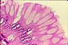
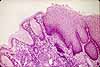
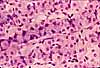
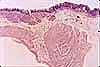
Thin section of the stomach's mucosal glands. This specimen was prepared by embedding in plastic rather than paraffin and slicing at 2µm thickness vs. 5-6 µm for routine sections.
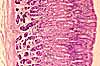
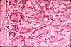
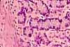
This preparation provides adequate resolution to see details of both parietal cells (P) and chief cells (C) as they appear in the deep portion of gastric glands.
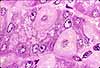
Secretory vesicles (containing pepsinogen) are clearly visible in the apical cytoplasm of chief cells.
Within the cytoplasm of parietal cells, relatively lightly-stained regions suggest the presence of intracellular canaliculi (into which acid is pumped).
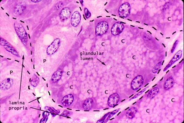
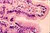
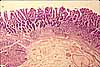
The epithelial nature of the chief cells can be readily seen in this section, which cuts across a gastric gland to reveal both the glandular lumen and also the surrounding lamina propria. Dashed black lines mark the boundary between gland and lamina propria.
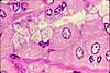
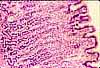
The tubular gastric glands tend to be rather twisted deep in the mucosa, so that neat cross sections may occasionally be found even in sections perpendicular to the mucosal surface. More commonly, however, the plane of section passes tangentially or obliquely through the cells which comprise the wall of the gland. Thus, at low magnification, gastric glands often appear cord-like rather than tubular.Contrast the thin-section appearance of these chief cells and parietal cells with that of other secretory epithelial cells in the gastric mucosa:
Related examples:
 |
 |
 |
 |
 |
 |
 |
 |
 |
 |
 |
Comments and questions: dgking@siu.edu
SIUC / School
of Medicine / Anatomy / David
King
https://histology.siu.edu/erg/GI136b.htm
Last updated: 10 May 2022 / dgk