
Notes
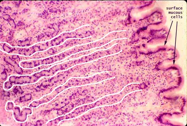
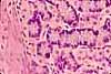
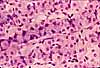
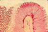
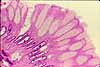
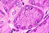
Most of the bulk of the gastric mucosa is occupied by secretory cells of the gastric glands, primarily parietal cells and chief cells, together with lamina propria.
In routine sections, these cells often appear to form cord-like bands of cells rather than distinct tubules. These represent glands sliced tangentially, with the plane of section passing through the cells which comprise the gland's wall without revealing the lumen.
Several such glands are outlined (with considerable artistic license) in the image above. Although every gland opens into a gastric pit (with several glands per pit), continuity of glandular epithelium with the surface epithelium is seldom evident in routine sections.
Cells with basally basophilic cytoplasm and basal nuclei are the chief cells. These are concentrated in the deeper regions of the glands, toward the left side of this image.
Cells with conspicuous eosinophilic cytoplasm and centrally located nuclei (not infrequently, two nuclei in one cell) are the parietal cells. These are concentrated in the middle to upper regions of the glands, across the middle of the image.
The glands are separated by thin strands of lamina propria, containing the denser, flattened or irregular nuclei of fibroblasts, capillary endothelium, and various cells of the immune system.
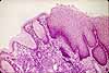
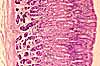
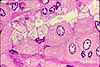
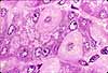
Related examples:
 |
 |
 |
 |
 |
 |
 |
 |
 |
 |
 |
 |
 |
 |
Comments and questions: dgking@siu.edu
SIUC / School
of Medicine / Anatomy / David
King
https://histology.siu.edu/erg/GI091b.htm
Last updated: 10 May 2022 / dgk