
Liver, overview
Notes
Click anywhere on this image to view the micrograph with labels.
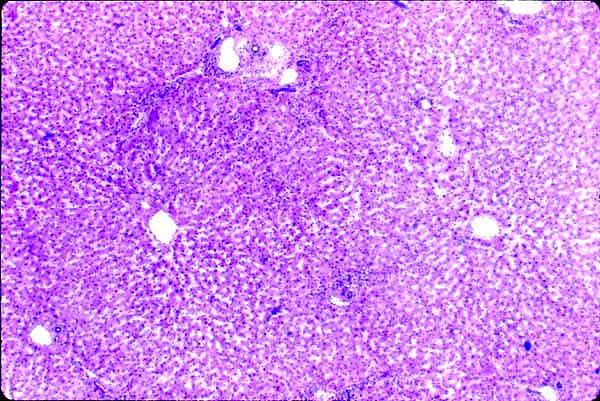
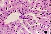
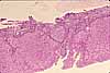
The liver appears, at first glance, like a rather unorganized mass of cells. But with a little care, one can detect the lobular organization of this tissue.
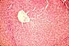
To visualize lobules, first locate several portal areas. These are readily recognizable as small patches of connective tissue, each containing a duct, a large vein, and a small artery. These mark the corners where lobules come together. (View a portal area.)
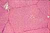
Then look for central veins. These are conspicuous spaces, with no associated connective tissue, located roughly midway between portal areas. These central veins mark the centers of lobules. (View a central vein.)
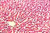
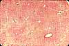
Lobules appear much more clearly in pig liver, which has an envelope of fibrous connective tissue around each lobule. (This tough connective tissue is one reason why pig liver, unlike calf liver or chicken liver, is not a popular menu item.)
Related examples:
|
|
 |
|
|
 |
 |
 |
 |
 |
 |
 |
 |
 |
 |
 |
 |
Comments and questions: dgking@siu.edu
SIUC / School
of Medicine / Anatomy / David
King
https://histology.siu.edu/erg/GI159b.htm
Last updated: 14 May 2022 / dgk