
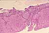
Liver, portal area
Notes
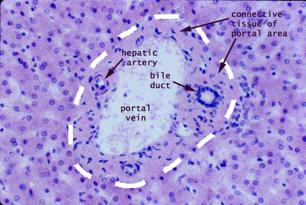
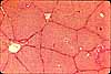
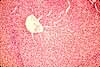
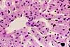
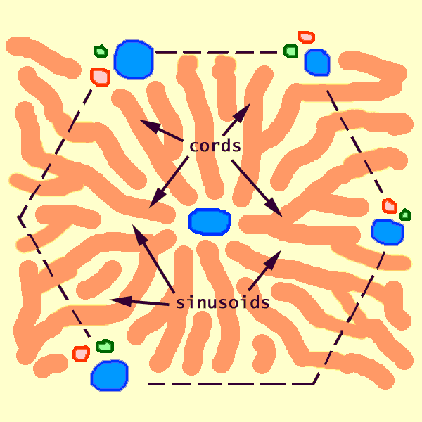
Portal areas (also called portal triads or portal canals) are located at the corners of liver lobules. Portal areas are normally surrounded by much larger areas packed with hepatic cords and sinusoids.
Each portal area contains three (hence the term portal triad) more-or-less conspicuous tubular structures all wrapped together in connective tissue.
- a branch of the bile duct
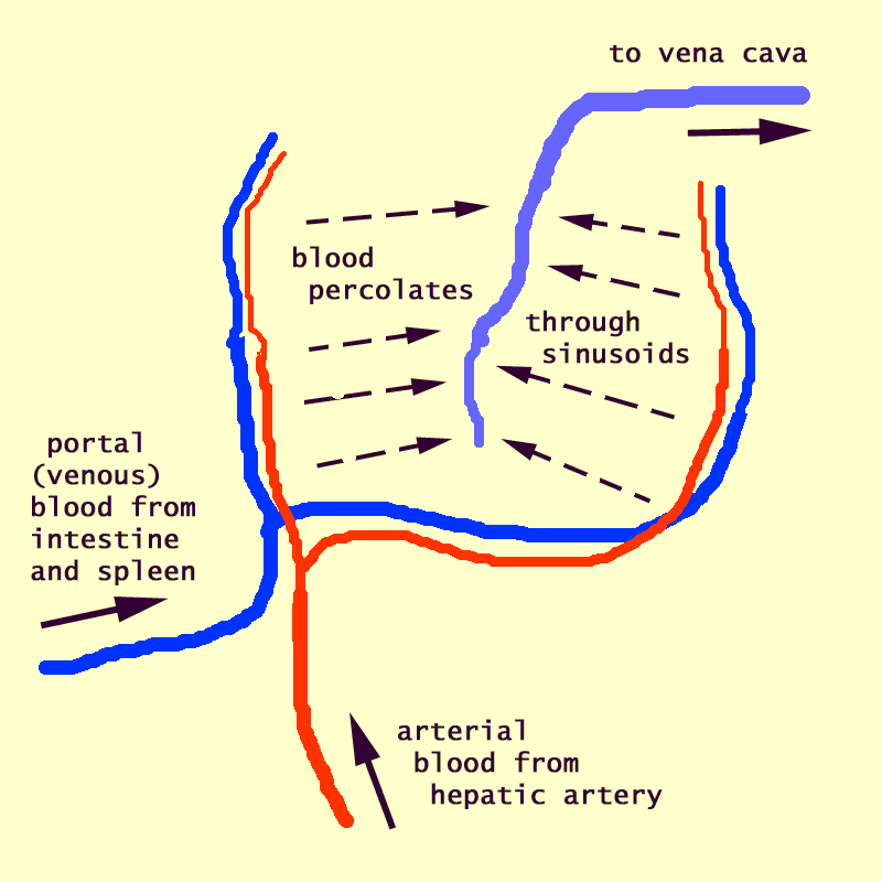
- a branch of the portal vein
- a branch of the hepatic artery
Of these three, the bile duct is the most easily recognized, by the conspicuous round nuclei in its cuboidal epithelium.
(A typical portal area also contains two additional structures, a nerve and a lymphatic channel, but these are seldom apparent in routine specimens.)
The portal areas visible in any random section of liver vary widely in the absolute sizes of the vessels and ducts therein.
Since the portal vein brings much more blood to the liver than does the hepatic artery, each branch of the portal vein is typically much larger than the associated branch of the hepatic artery. The relative sizes of the paired vessels in a portal area thus differ from those of a typical vein / artery pair in other parts of the body, where the artery delivers the same volume of blood that the vein subsequently returns.
An increase in the amount of connective tissue extending out from portal areas is characteristic of cirrhosis (sclerosis / fibrosis / scarring of liver), in which damaged hepatic tissue is replaced by scar tissue (fibrous connective tissue). For images of cirrhosis, see WebPath or Milikowski & Berman's Color Atlas of Basic Histopathology, pp. 284ff.
Related examples:
 |
 |
 |
|
 |
 |
 |
 |
 |
 |
 |
 |
 |
 |
 |
 |
 |
Comments and questions: dgking@siu.edu
SIUC / School
of Medicine / Anatomy / David
King
https://histology.siu.edu/erg/GI162b.htm
Last updated: 14 May 2022 / dgk