

Notes
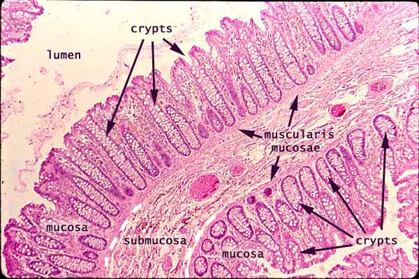
The mucosa of the colon is characterized by straight crypts with no villi.
In this specimen, the plane of section passes through a wrinkle in the wall, so that submucosa passes diagonally across the image with mucosa appearing on both sides.
When crypts are cut longitudinally (as in much of the mucosa in the upper portion of this image), the columns of lamina propria between the epithelium of adjacent crypts might be misinterpreted as villi. But note that the lumenal spaces within the crypts are uniformly narrow (when they are visible at all), not variable and often wide, as spaces between villi of the small intestine.
Also note that when the plane of section cuts across a crypt, the crypt appears to be surrounded by lamina propria. In contrast, when villi are cut across, they appear as isolated islands in the lumen.
Note the large number of goblet cells (clear "bubbles") in crypt epithelium.
The prominent pink ovals in the submucosa are blood vessels filled with red blood cells.
Related examples:
Comments and questions: dgking@siu.edu
SIUC / School
of Medicine / Anatomy / David
King
https://histology.siu.edu/erg/GI182b.htm
Last updated: 17 May 2022 / dgk