Notes
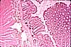
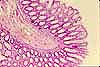
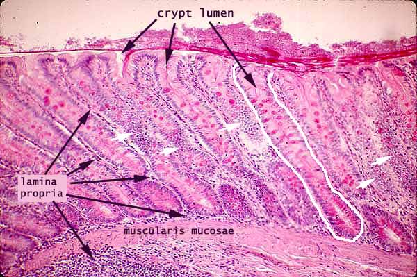
The mucosa of the colon is characterized by straight crypts with no villi. One crypt is outlined in white.
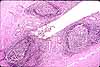
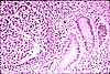
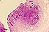
The appearance of lamina propria beneath the muscularis mucosae indicates that the plane of section has cut through a wrinkle in the mucosa. That this layer is not submucosa is indicated by the large number of lymphocyte nuclei. The apparent thickness of the muscularis mucosae also suggests that it has been cut tangentially, as does its disappearance at the lower left corner of the image.
This specimen has been stained with PAS (per-iodic acid, Schiff), so that mucus in goblet cells appears red.
Note how many epithelial nuclei are visible where crypt epithelium has been cut tangentially (white arrows). Most of these nuclei belong to absorptive cells, which far outnumber the goblet cells in colon epithelium (as also in the small intestine), even though they are crowded by goblets and hence inconspicuous.
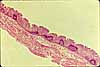
In routine H&E preparations, the mucus in goblet cells appears as clear "bubbles" in crypt epithelium.
Related examples:
Comments and questions: dgking@siu.edu
SIUC / School
of Medicine / Anatomy / David
King
https://histology.siu.edu/erg/GI142b.htm
Last updated: 27 May 2022 / dgk