Penis
From the perspective of histology, the most remarkable feature of the penis is its core of erectile tissue.
 Penile erectile tissue is organized into paired dorsal corpora cavernosa and
one ventral corpus spongiosum. The corpora cavernosa are
surrounded by tough fibrous connective tissue, the tunica albuginea. Between
this sheath and the overlying skin is a layer of very loose elastic connective
tissue that permits the skin of the penis to move freely along
the shaft. The
penile urethra passes through the corpus spongiosum, where it
is associated with small mucous glands of Littre.
Erectile tissue contains a specialized arrangement of arteries, shunts, and anastomosing sinusoids within a matrix of connective tissue and smooth muscle, forming "a highly structured criss-crossing of interconnected fibers and spaces that are tensed as the cylinder expands during erection. This creates an internal strength and rigidity that is far greater than that possible in a hollow tube filled to equivalent pressure" (quoted from Barreto, Caballer, and Cubilla, in Sternberg's Histology for Pathologists, 2nd. ed., p. 1045).
Arterial blood entering the central artery of each cavernous body may flow either into an arteriovenous shunt or into a network of trabecular vessels. In a normally flaccid penis, the shunt is dilated so that most blood passes through the dilated shunt. During erection, vasoconstriction reduces the opening into the shunt, directing blood into the trabecular spaces which are thereby inflated with blood. (You might imagine the result as somewhat like a bundle of very many long, narrow balloons. When inflated, such a bundle can become quite stiff.) During erection, the corpus spongiosum protects the urethra from compression by the engorged corpora cavernosa.
For a much more thorough verbal description of the penile anatomy, see male genital anatomy at Boston University School of Medicine. For a much better image than can be found in this website, see Histology Guide.
For the role of fibroblasts in penile erection, see Science 383 (9 Feb 2024), DOI: 10.1126/science.ade8064.
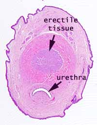
 The slide in our reference sets (e.g., the image at right) may not match this
description, presumably because it represents a very immature organ (note
the specimen's actual size on its microscope slide), in which the erectile tissue is not yet fully developed. Nevertheless,
you should be able to observe several prominent features including erectile
tissue of the corpora cavernosa with numerous interconnected vascular channels,
the surrounding dense connective connective tissue of the tunical albuginea,
conspicuous nerves and blood vessels, the urethra lined by transitional epithelium
(urothelium), and keratinized epithelium of the skin.
For additional images of erectile tissue and other specialized penile tissues, consult Sternberg's Histology for Pathologists.


