

Notes
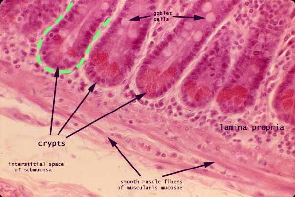
In this view of the jejunum, the mucosa is at the top and the submucosa is at the bottom.
Individual smooth muscle fibers, comprising the muscularis mucosae, define the boundary between mucosa and submucosa.
Crypts are a prominent feature in the mucosa.
One crypt is outlined in green. The crypt lumen is visible at the center of this crypt.
Bright red color near the base of each crypt shows the location of secretory granules of Paneth cells. (These granules are not conspicuous in most H&E specimens.)
Pale "bubbles" in the upper portion of the crypts are mucous droplets in goblet cells.
The connective tissue between and beneath the crypts is lamina propria.
The many small nuclei in lamina propria are mostly lymphocytes. (Identifying individual cells in lamina propria is not practical in most routine specimens.)
More small intestine examples:
 |
 |
 |
 |
 |
 |
 |
 |
 |
 |
 |
 |
 |
 |
 |
 |
 |
 |
 |
 |
 |
 |
Comments and questions: dgking@siu.edu
SIUC / School
of Medicine / Anatomy / David
King
https://histology.siu.edu/erg/GI007b.htm
Last updated: 22 July 2023 / dgk