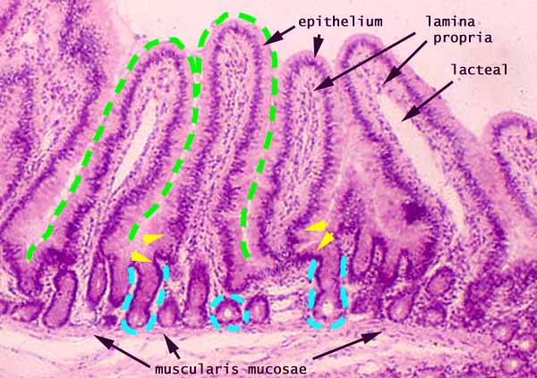

Small Intestine, mucosa (cross section of jejunum)

Notes
(View this image without marks and labels.)
The mucosa of the small intestine is lined by simple columnar epithelium which evaginates into villi (green outlines) and invaginates into crypts (blue outlines).
Note that epithelium of villi is continuous with that of adjacent crypts (yellow arrowheads), even though some crypts appear "disconnected" due to plane of section.
Lamina propria of the small intestine forms the core of villi and surrounds the crypts. A long "empty" space within the lamina propria, especially if lined by simple squamous endothelium, is a lacteal.
The muscularis mucosa of the small intestine forms a thin layer (only a few muscle fibers in thickness) beneath the deep ends of the crypts.
More small intestine examples:
 |
 |
 |
 |
 |
 |
 |
 |
 |
 |
Comments and questions: dgking@siu.edu
SIUC / School
of Medicine / Anatomy / David
King
https://histology.siu.edu/erg/GI031b1.htm
Last updated: 27 May 2022 / dgk