
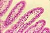
Small Intestine, shapes of villi
Notes
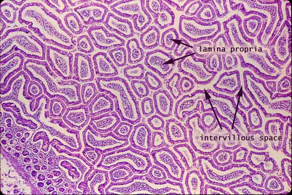
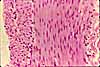
The cross-sectional shapes of villi can be plainly seen when the intestinal mucosa is cut tangentially, as in this image.
Each villus is outlined by a ring of epithelium surrounding a core of lamina propria. (Note that the space between the epithelium and the lamina propria is a fixation artifact, resulting from shrinkage of the tissue during preparation.)
Some villi have a finger-like shape, while others are more flattened. The length and shape of of villi changes somewhat along the length of the intestine, with villi tending to be flatter in the duodenum and longer and more finger-like in the jejunum.
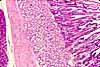
Crypts (small epithelial circles, surrounded by lamina propria) and muscularis mucosa appear at the lower left.
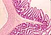
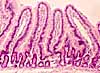
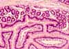
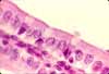
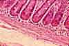
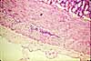
More small intestine examples:
 |
 |
 |
 |
 |
 |
 |
 |
 |
Comments and questions: dgking@siu.edu
SIUC / School
of Medicine / Anatomy / David
King
https://histology.siu.edu/erg/GI037b.htm
Last updated: 27 May 2022 / dgk