 Notes
Notes
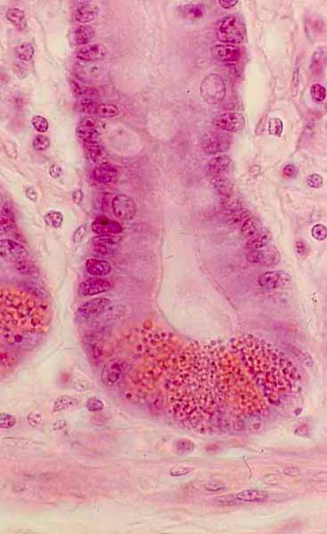
Intestinal crypt, with Paneth cells
 Notes
Notes
Click on the image to identify the tissues. (Depending on your browser, simply "hovering" over an area may produce a label.)
Crypts are a prominent feature of the intestinal mucosa.
The simple columnar epithelium which lines the crypt consists of stem cells and newly-formed absorptive and goblet cells.
At the bottom of the crypt are Paneth cells. Each paneth cell has a typical serous cell appearance, with secretory vesicles (bright red in this micrograph) containing lysosomal enzymes packed into apical cytoplasm. (These granules are not conspicuous in most H&E specimens.)
The many small nuclei in lamina propria are mostly lymphocytes. (Identifying individual cells in lamina propria is not practical in most routine specimens.)
Related examples:
 |
 |
 |
 |
 |
 |
 |
 |
 |
Comments and questions: dgking@siu.edu
SIUC / School
of Medicine / Anatomy / David
King
https://histology.siu.edu/erg/GI112b.htm
Last updated: 17 May 2022 / dgk