
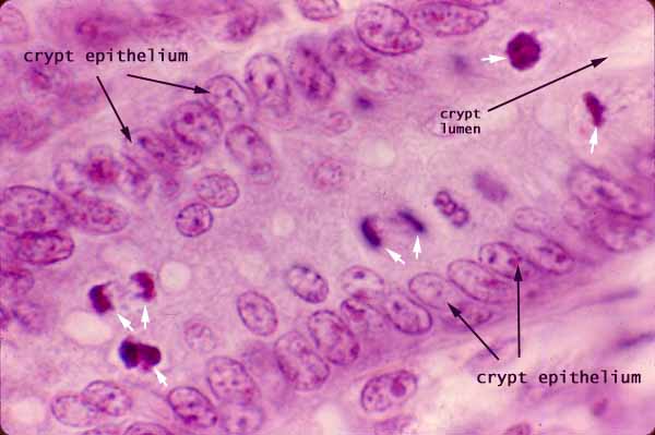
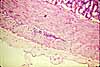
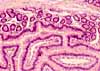
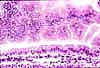
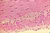
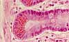
Small Intestine, dividing cells in crypt
Notes
The absorptive cells and goblet cells of the small intestine have a short life-span (a few days) and are continually replaced by dividing stem cells in the crypts. Evidence of this cell-replacement activity can often be seen in the form of mitotic figures (white arrows, above).
Mitotic figures are characterized by condensed chromosomes, which are intensely basophilic. The appearance of mitotic figures varies with the stage of mitosis (prophase, metaphase, anaphase, telophase) and with the orientation of the dividing cell relative to the plane of section.
The nucleus of a columnar epithelium cell migrates toward the cell's apical end prior to mitosis, so that mitotic figures and nuclei of recently-divided cells typically occur closer to the lumen than the resting nuclei.
Lamina propria appears in the upper left and lower right corners of this image.
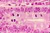
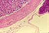
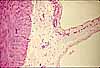
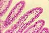
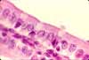
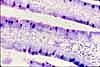
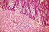
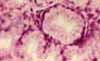
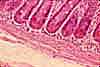
More small intestine examples:
 |
 |
 |
 |
 |
 |
 |
 |
 |
 |
 |
 |
 |
 |
 |
 |
 |
 |
|
|
 |
 |
 |
Comments and questions: dgking@siu.edu
SIUC / School
of Medicine / Anatomy / David
King
https://histology.siu.edu/erg/GI038b.htm
Last updated: 27 May 2022 / dgk