Topics for today:
- Website for Lincoln Scholars Program Histology
Just Google "Lincoln Histology" or "SIU Histology".
- Basic tissue types
- Connective tissue
- Lung
- Inflammation (as time allows)
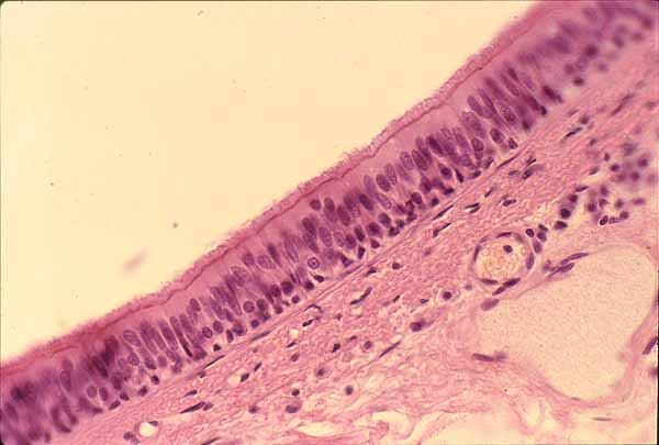
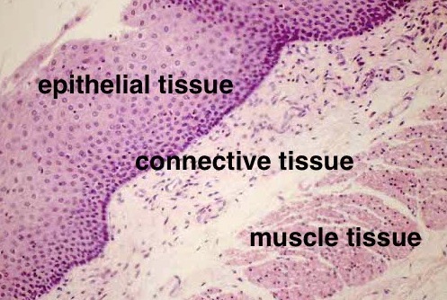
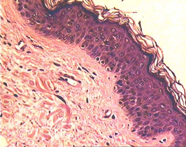
What follows are very sketchy notes. Follow the links above for more thorough presentation of the topics below. Thumbnail images below are also links to additional information.Four basic tissue types: nervous, muscle, epithelial, connective. (Here only epithelial and connective.)
Epithelium sits on top of connective tissue.
Connective tissue is the stuff which sits immediately underneath epithelium.
Most of the time, epithelial cells form surfaces.
All of the time, epithelial cells are attached to one another.
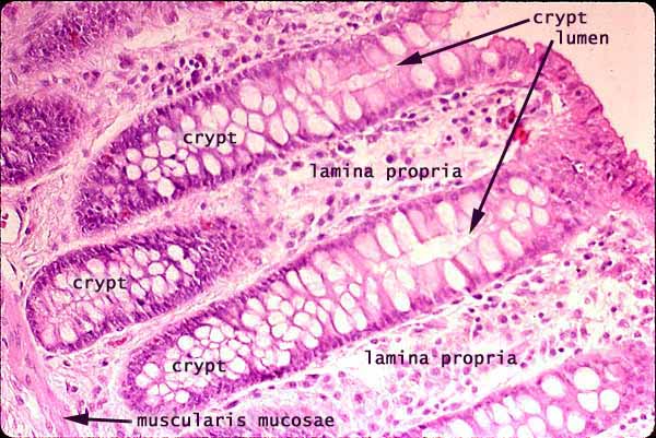
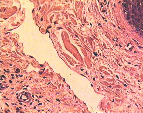
colors of tissues in micrographs
Most tissue components are colorless. The colors you see in routine micrographs are from dyes added for contrast. The most commonly used dyes are hematoxylin (deep purple or blue) and eosin (pink, named after Eos, the "rosy fingered goddess of the dawn"). Collagen stains pink with eosin. But not all pink stuff is collagen. Muscle fibers also stain pink with eosin. Cytoplasm sometimes stain pink, usually tinged with purple. But since collagen is the most abundant protein in the body, in many micrographs most of the pink stuff will be collagen. Cell nuclei typically as deeply-stained dark dots (at low magnification).
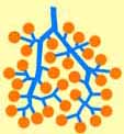
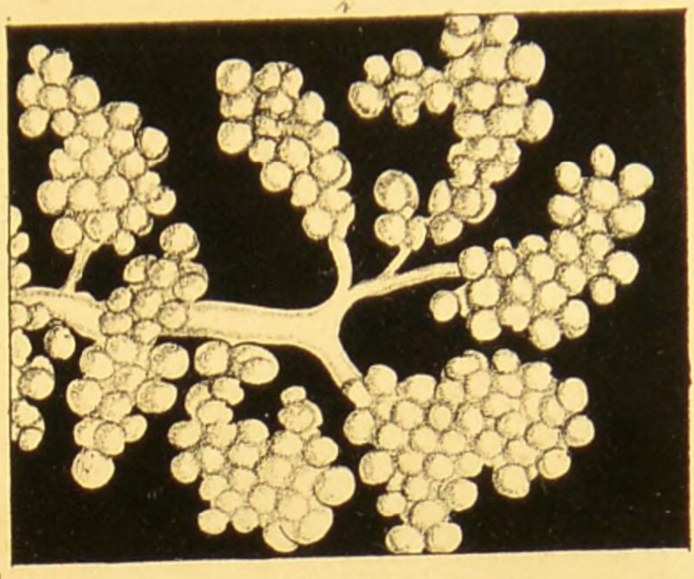
glands
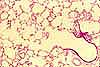
A gland may be shaped like a bunch of grapes.


Connective tissue consists of an assortment of stuff: There are cells. There are fibers. There is ground substance.
Connective tissues varies from place to place, with significant differences in the proportion of the various components. Blood and bone are extreme examples.
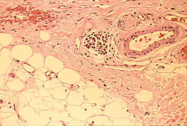
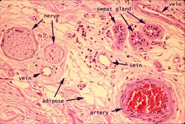
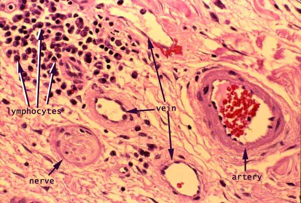
Nerves and blood vessels travel through connective tissue, are surrounded by connective tissue.
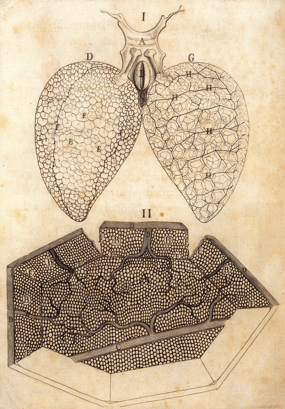
Epithelial tissue is inconspicuous in lung alveoli. Each thin wall between alveoli is a sandwich, with very thin flat epithelial cells on either side of connective tissue containing capillaries.
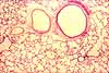
The underlying tissue organization of lung is quite similar to that of a gland, where the "bunch of grapes" is inflated with air and all the tissues squeezed into the thin walls between alveolar walls.
grapes, again
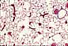
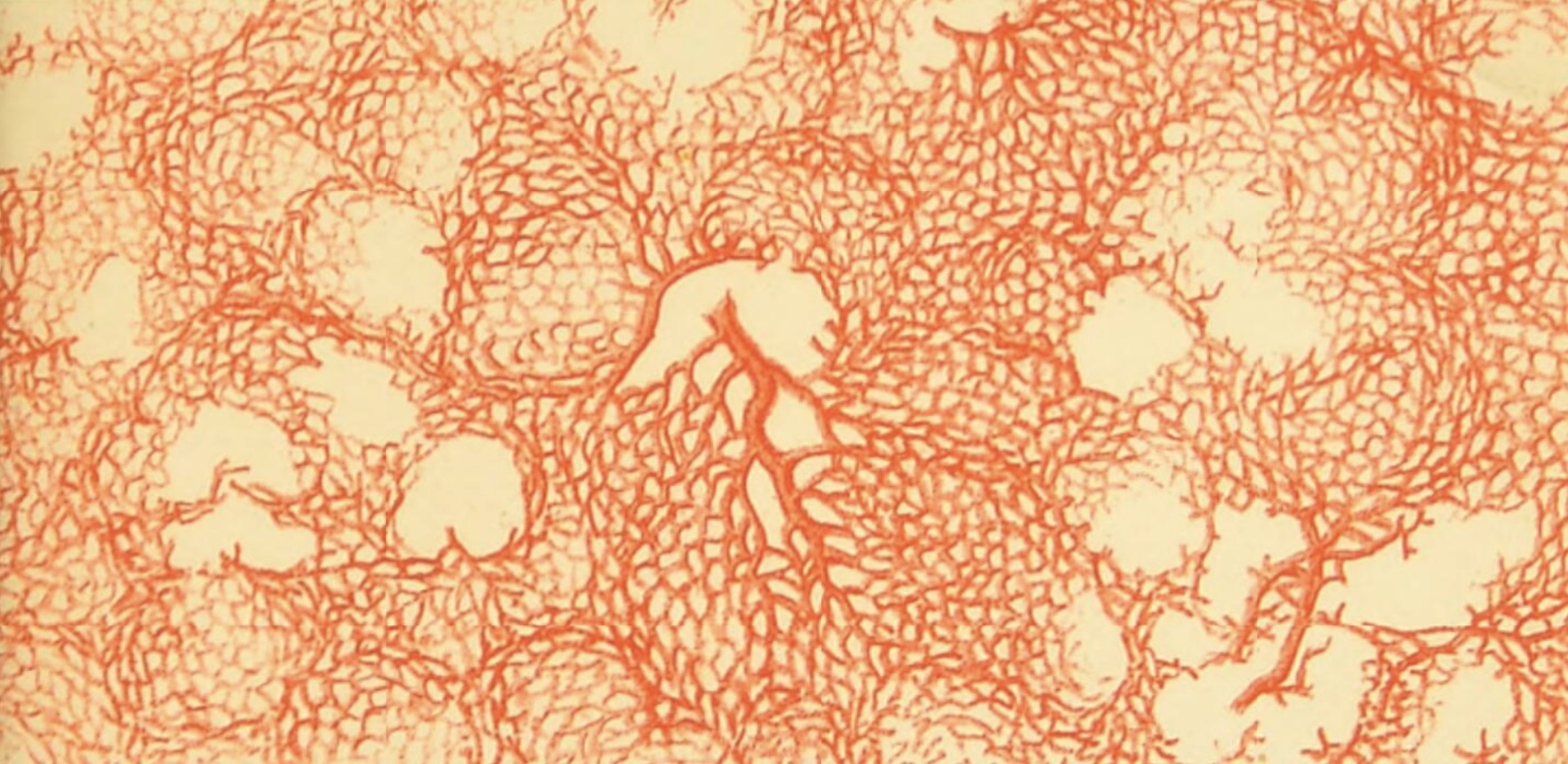
capillaries of lung
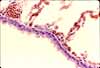
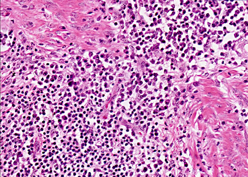
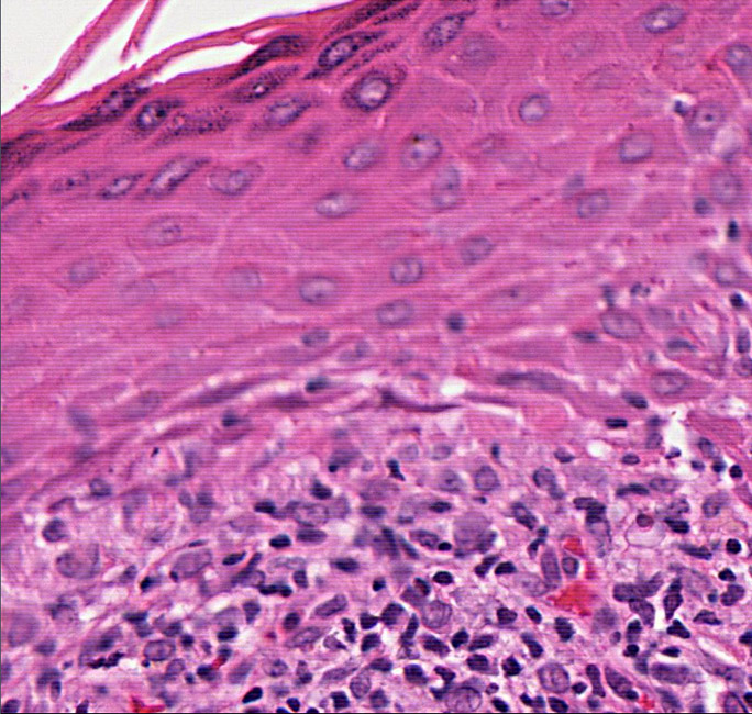
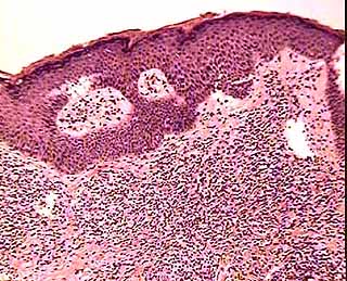
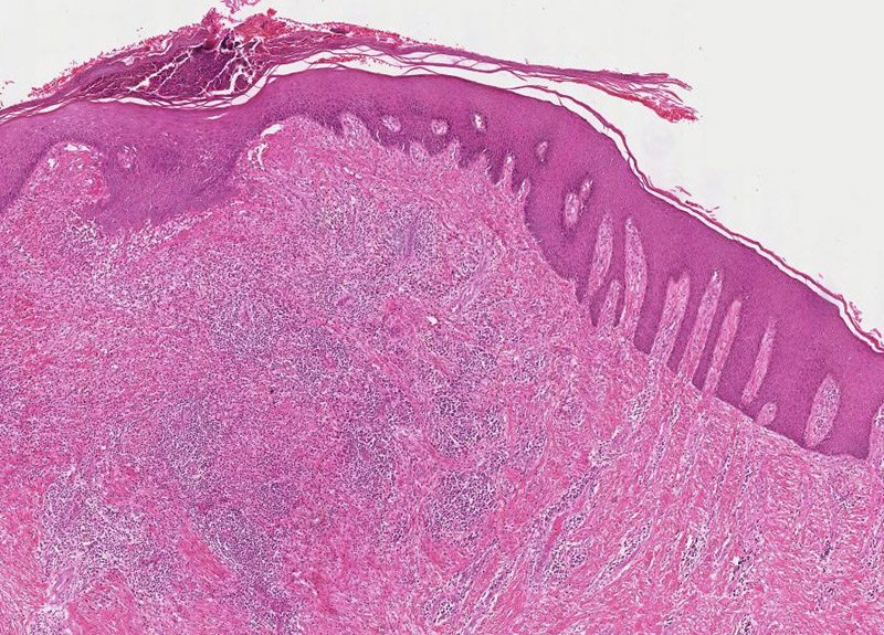
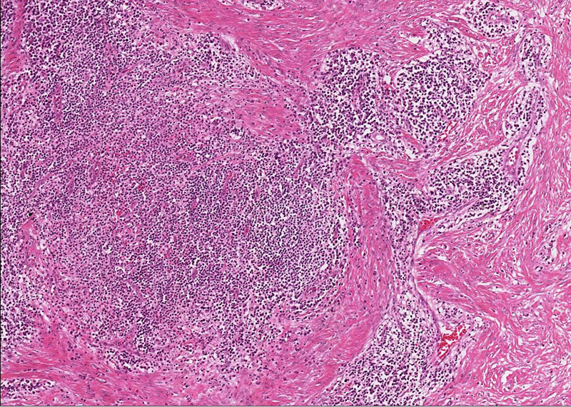
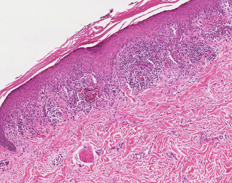
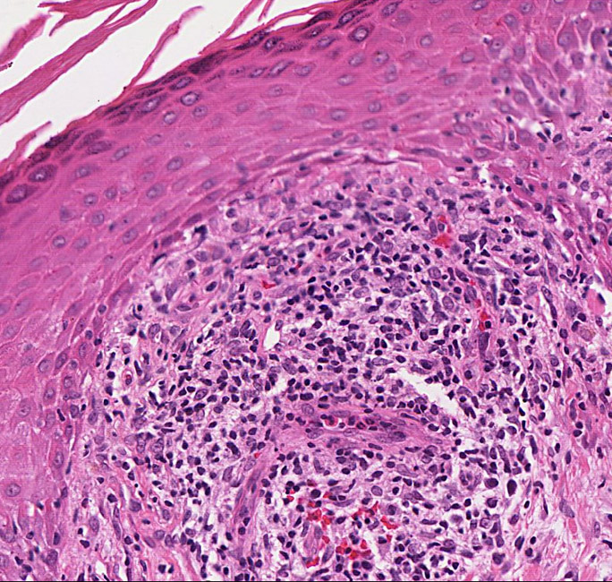
Inflammation is recognizable in tissue specimens as the presence if very many small cell nuclei in connective tissue, the inflammatory infiltrate of neutrophils and lymphocytes. (check time)
examples of inflammation in skin
examples of inflammation in lung
- Contents for traditional curriculum
- Image index for this website
- If you have a taste for history, see Eponyms in histology.
SEARCH THE SITE:
Comments and questions: dgking@siu.edu
SIUC / School of Medicine / Anatomy / David King
https://histology.siu.edu//LincolnScholarsProgram/15Aug.htm
Last updated: 11 June 2023 / dgk