
Histology Topics for Lincoln Scholars ProgramElementary histopathology.
- Introductory comments
- Illustrative examples
- Index to examples
- Some basic vocabulary
- Special instructions for viewing examples
- Examples for comparing normal with pathological histology
- List of linked WebPath images
- Additional resources for histopathology
♥ Inflammation, cell death, tissue repair, and scarring, and cancer are all fundamentally tissue-level phenomena.
To understand these basic pathological processes, knowledge of normal histology is essential.
Each of these pathological processes yields readily recognizable signs, IF normal tissue architecture is already familiar.
- Inflammation is indicated by the presence of numerous infiltrating leukocytes.
Inflammation presents as lots of small nuclei (lymphocytes and/or neutrophils) gathered in places where such gatherings are not typical. (Lymphoid tissues, such as tonsils and appendix, can normally offer a similar appearance.)- Cell death can be indicated by condensation of nuclei (pyknosis), fragmentation of nuclei (karyorrhexis), and loss of nuclei. (For images of pyknosis in normal tissue, see sebaceous glands.)
- Scarring and/or fibrosis is indicated by collagen in amounts and/or locations where it is not expected.
- Cancer is indicated by increased amounts and disturbed patterns of tissue growth.
Cellular signs of malignancy include:
- unusual variation in cell size and shape;
- abnormal variation in size and shape of nuclei;
- increased numbers of mitotic cells,
- bizarre mitotic figures.
See examples below, under "neoplasia," for images of malignant and metastatic neoplasm.Problem cases in our curriculum might not engage these topics at the tissue level until your second or third year. However, experience teaches that early exposure to any topic will ease the way into more intensive engagement that may come later.
This page offers a number of examples contrasting normal tissue appearance with pathology images selected from The Internet Pathology Laboratory for Medical Education website (WebPath). These examples -- all of which should be straightforward for beginners to appreciate -- are intended to motivate and encourage study of normal histology early in your medical-school experience (i.e., before the need to understand histopathology becomes urgent).
INDEX OF EXAMPLES
|
ANATOMICAL LOCATION |
Inflammation
|
Metaplasia
|
Dysplasia
|
Neoplasia
|
Scarring and/or
Fibrosis |
Cell death
|
|
Liver
|
||||||
|
Cervix
|
||||||
|
Salivary
gland |
||||||
|
Esophagus
|
||||||
|
Small
intestine |
||||||
|
Appendix |
||||||
|
Colon
|
||||||
|
Lung
|
||||||
|
Nasal
mucosa |
||||||
|
Larynx
|
||||||
|
Heart
|
||||||
|
Testis
|
||||||
|
Prostate
|
||||||
|
Uterus
|
||||||
|
Kidney
|
Some vocabulary, helpful for reading descriptions of pathology:
- Parenchyma and stroma are convenient terms to distinguish between principal tissue components of an organ:
- Parenchyma refers to the distinctive cell types which conduct the specific function of an organ, or the distinctive cell types which characterize a tumor.
- Stroma is tissue which supports the parenchyma -- connective tissue, blood vessels, nerves.
- Inflammation is a fundamental mechanism of tissue defense.
Inflammation accompanies many different types of tissue disturbance.
The suffix "-itis" indicates inflammation.
- Metaplasia is replacement of differentiated cells within a tissue by differentiated cells of a different type.
- Dysplasia is disordered growth of a tissue.
Dysplasia is commonly a prelude to malignant neoplasm.
- Neoplasia literally means "new growth."
The suffix "-oma" commonly indicates a neoplastic growth, or "tumor."
Neoplasms may be:The name of a neoplasm commonly indicates its tissue-type of origin. For example:
- benign -- confined in location,
- malignant -- invading into surrounding tissue
- metastatic -- disseminating to distant locations.
- A "carcinoma" is a malignant neoplasm of epithelial origin.
- An "adenoma" is a tumor of glandular origin.
- Scarring is replacement of damaged tissue by collagen.
- Fibrosis is a more general term, indicating excess deposition of collagen for any reason.
- (The terms above are far from a complete list of histopathology vocabulary.)
Illustrative examples
Each example below includes both an image of normal histology and a link to a WebPath image from a similar anatomical location, illustrating pathologically altered histology.
The WebPath links will take you to pages at WebPath's General Pathology or Systemic Pathology collections. In addition to a micrograph of the pathology, each of these links offers a brief description of the illustrated pathology as well as further links to related WebPath resources.
NOTE: These examples are intended for beginners. Many of WebPath links include pathological tissue alongside normal within the same image, so that the difference should be apparent even if you have not yet learned much about normal tissue appearance.
Special instructions for viewing these examples:
- Within each example, the normal image is itself a link to descriptive information about the tissue illustrated.
- Within each example, the WebPath link will open a new tab at the WebPath website.
Once opened, each WebPath page will easily link to related WebPath examples.
- To compare normal with pathological appearance, toggle back and forth between the two tabs.
- Alternatively, you might open this page in two adjacent windows.
- Adjust the width of these two windows so that they fit side-by-side on your screen
[presuming your computer monitor has sufficient width].- Keep both windows open.
- One window will display each normal image and its associated comment.
- The other window will display each WebPath link, in parallel with the corresponding normal image.
- When comparing images, pay attention not to color (staining quality can vary tremendously from one preparation to another) but to tissue architecture -- the structure and arrangement of cells.
- Close the WebPath tab when you are ready to move to another example: Otherwise tabs will accumulate indefinitely in your browser window.
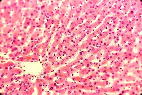
Example 01
Image above: Normal liver parenchyma
WebPath example: Malignant neoplasm in liver. [Link opens in new tab.]
In the WebPath image, normal liver parenchyma appears on left, malignant tissue on right.REMINDER: Close tab for WebPath example.
TOP OF PAGE / INDEX OF EXAMPLES / BASIC VOCABULARY
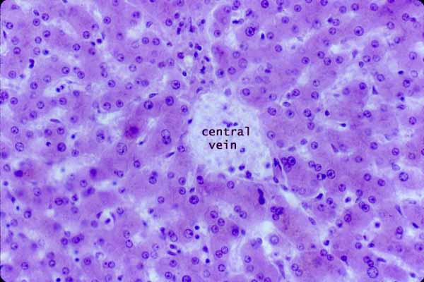
Example 02
Image above: Normal liver parenchyma
WebPath example: Hepatitis. [Link opens in new tab.]
In the WebPath image, a swath of inflammatory infiltrate appears in a band of connective tissue from lower left to upper right. Tissue at upper left is reasonably normal hepatic parenchyma.REMINDER: Close tab for WebPath example.
TOP OF PAGE / INDEX OF EXAMPLES / BASIC VOCABULARY
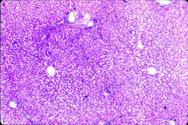
Example 03
Image above: Normal liver parenchyma (low magnification)
WebPath example: Cirrhosis (scarring of liver). [Link opens in new tab.]
In the WebPath image, two patches of hepatic parenchyma (surviving remnants of liver lobules) are surrounded by broad bands of connective tissue. This scarring replaces parenchyma that had been previously destroyed.REMINDER: Close tab for WebPath example.
TOP OF PAGE / INDEX OF EXAMPLES / BASIC VOCABULARY
Example 04
Image above: Normal liver parenchyma
WebPath example: Malignant neoplasm in liver. [Link opens in new tab.]
In the WebPath image, normal liver parenchyma appears on right, malignant tissue on left.REMINDER: Close tab for WebPath example.
TOP OF PAGE / INDEX OF EXAMPLES / BASIC VOCABULARY
Example 05
Image above: Normal liver parenchyma
WebPath example: Malignant neoplasm in liver. [Link opens in new tab.]
In the WebPath image, normal liver parenchyma appears on left, malignant tissue on right.REMINDER: Close tab for WebPath example.
TOP OF PAGE / INDEX OF EXAMPLES / BASIC VOCABULARY
Example 06
Image above: Normal vagina (cervical epithelium is similar)
WebPath example: Chronic cervicitis. [Link opens in new tab.]
In the WebPath image, note numerous small dark nuclei (lymphocytes) in connective tissue beneath the epithelium, as well as extravasated red blood cells (red color).REMINDER: Close tab for WebPath example.
TOP OF PAGE / INDEX OF EXAMPLES / BASIC VOCABULARY
Example 07
Image above: Normal vagina (cervical epithelium is similar)
WebPath example: Cervical dysplasia. [Link opens in new tab.]
In the WebPath image, normal cervical epithelium is at left, dysplastic epithelium at rightn.REMINDER: Close tab for WebPath example.
TOP OF PAGE / INDEX OF EXAMPLES / BASIC VOCABULARY
Example 08
Image above: Normal vagina (cervical epithelium is similar)
WebPath example: Cervical dysplasia with inflammation. [Link opens in new tab.]
In the WebPath image, inflammatory infiltrate underlies the abnormally thickened dysplastic epithelium (at right) but not the normal epithelium (at left). The thin dark lines crossing the epithelium are artefactual wrinkles in the tissue section.REMINDER: Close tab for WebPath example.
TOP OF PAGE / INDEX OF EXAMPLES / BASIC VOCABULARY
Example 09
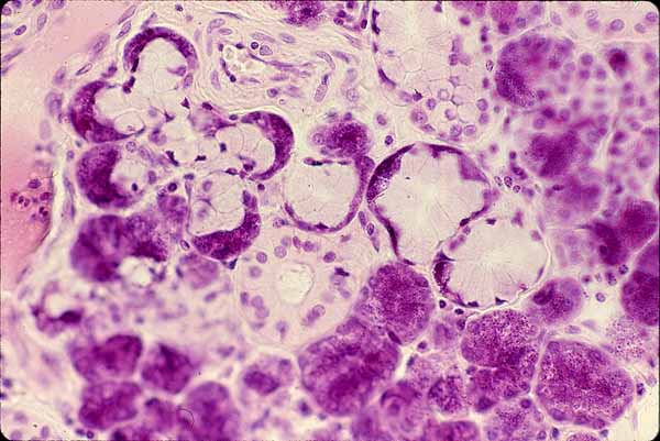
Image above: Normal salivary gland
WebPath example: Inflammation and fibrosis of salivary gland. [Link opens in new tab.]
In the WebPath image, extensive connective tissue (the "fibrosis) surrounds two patches of inflammatory infiltrate and several ducts. Remarkably little glandular parenchyma is present (i.e., few secretory cells). Connective tissue is normally an inconspicuous tissue component amidst the epithelial parenchyma of salivary glands.REMINDER: Close tab for WebPath example.
TOP OF PAGE / INDEX OF EXAMPLES / BASIC VOCABULARY
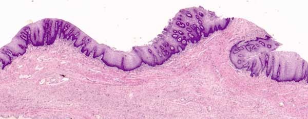
Example 10
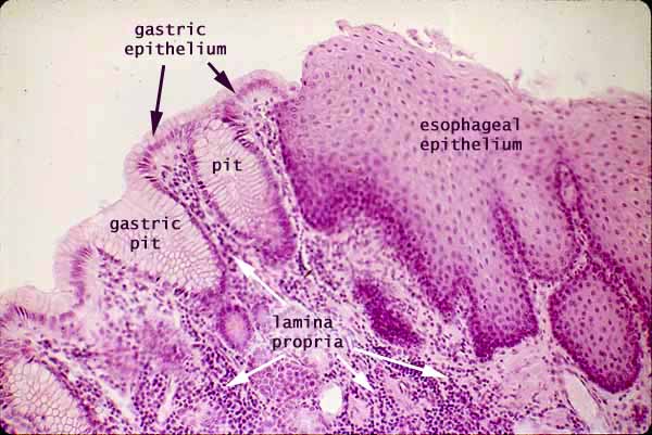
Image above: Normal gastro-esophageal junction
WebPath example: Esophageal metaplasia (Barrett's esophagus). [Link opens in new tab.]
In the WebPath image, although the transition from stratified squamous epithelium (at right) to simple columnar epithelium (at left) is superficially reminiscent of the normal gastro-esophageal junction, this transition lies within the esophagus proper (i.e., proximal to the normal site of epithelial transition). Furthermore, the metaplastic epithelium displays an intestinal character: Instead of a uniform epithelium of gastric surface mucous cells, the metaplastic columnar epithelium is interspersed with goblet cells, and this transformed epithelium forms crypt-like tubular invaginations rather than short gastric pits. Inflammatory infiltrate is seen in lamina propria.REMINDER: Close tab for WebPath example.
TOP OF PAGE / INDEX OF EXAMPLES / BASIC VOCABULARY
Example 11
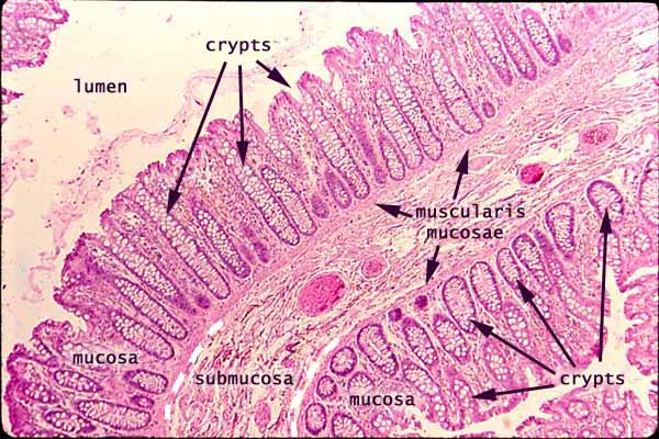
Image above: Normal colon mucosa
WebPath example: Adenomatous polyp in colon. [Link opens in new tab.]
In the WebPath image, the neoplasm ("new growth") is clearly distinct from surrounding normal mucosa, although of a similar character.REMINDER: Close tab for WebPath example.
TOP OF PAGE / INDEX OF EXAMPLES / BASIC VOCABULARY
Example 12
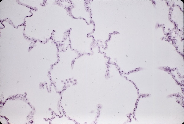
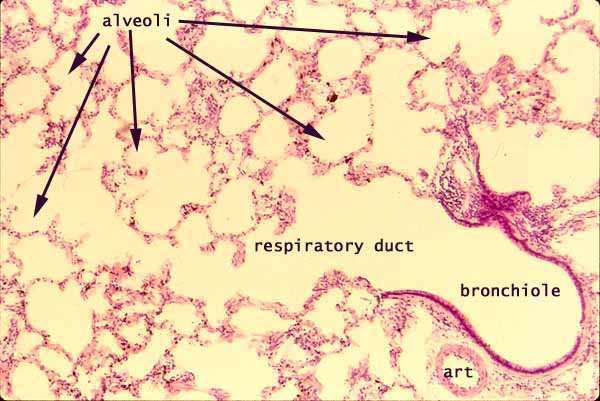
Image above: Normal lung alveoli
WebPath example: Pneumonia. [Link opens in new tab.]
In the WebPath image, alveolar lumens (which should contain only air) are filled with inflammatory cells and fluid.REMINDER: Close tab for WebPath example.
TOP OF PAGE / INDEX OF EXAMPLES / BASIC VOCABULARY
Example 13
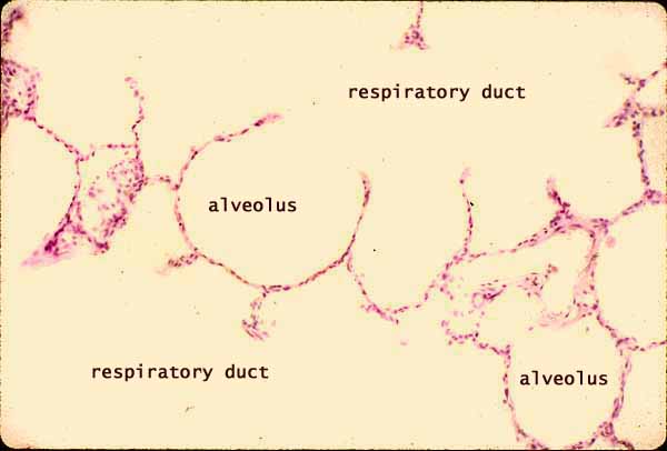
Image above: Normal lung alveoli
WebPath example: Pneumonia. [Link opens in new tab.]
In the WebPath image, alveolar lumens (which should contain only air) are filled with inflammatory cells and fluid.REMINDER: Close tab for WebPath example.
TOP OF PAGE / INDEX OF EXAMPLES / BASIC VOCABULARY
Example 14
Image above: Normal lung alveoli
WebPath example: Metastasis of breast cancer into lung. [Link opens in new tab.]
In the WebPath image, the conspicuous lump of basophilic tissue has no business appearing in lung. Alveolar lung tissue appears across the top of the WebPath image.REMINDER: Close tab for WebPath example.
TOP OF PAGE / INDEX OF EXAMPLES / BASIC VOCABULARY
Example 15
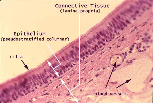
Image above: Normal respiratory epithelium
(This is tracheal epithelium; nasal epithelium is similar.)WebPath example: Anaphylaxis (allergic response) in nasal mucosa. [Link opens in new tab.]
In the WebPath image, normal ciliated columnar nasal epithelium appears at left; lymphocytes and eosinophils infiltrate the underlying connective tissue.REMINDER: Close tab for WebPath example.
TOP OF PAGE / INDEX OF EXAMPLES / BASIC VOCABULARY
Example 16
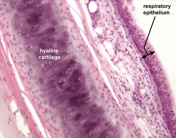
Image above: Normal respiratory epithelium
(This is tracheal epithelium; laryngeal epithelium is similar.)WebPath example: Squamous metaplasia of larynx. [Link opens in new tab.]
In the WebPath image, normal pseudostratified respiratory epithelium is at right, squamous metaplasia is at left.REMINDER: Close tab for WebPath example.
TOP OF PAGE / INDEX OF EXAMPLES / BASIC VOCABULARY
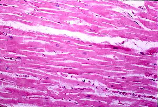
Example 17
Image above: Normal cardiac muscle
WebPath examples, Myocardial infarction [links open in new tabs]:
- WebPath "acute" (first day), inflammation not yet present.
- WebPath "recent", inflammation not yet present.
- WebPath 1-2 days, inflammation beginning.
- WebPath 3-4 days, acute inflammation established.
- WebPath 1-2 weeks.
- WebPath 3-5 weeks, with scarring.
- WebPath 2 months, with scarring.
REMINDER: Close tab for WebPath examples.
TOP OF PAGE / INDEX OF EXAMPLES / BASIC VOCABULARY
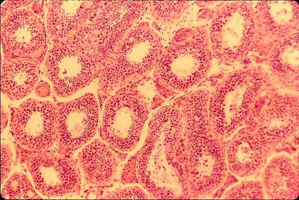
Example 18
Image above: Normal seminiferous tubules of testis
WebPath example: Seminoma. [Link opens in new tab.]
In the WebPath image, normal testis appears at left; at right are nests of neoplastic cells with dense inflammatory infiltrate in the surrounding stroma.REMINDER: Close tab for WebPath example.
TOP OF PAGE / INDEX OF EXAMPLES / BASIC VOCABULARY
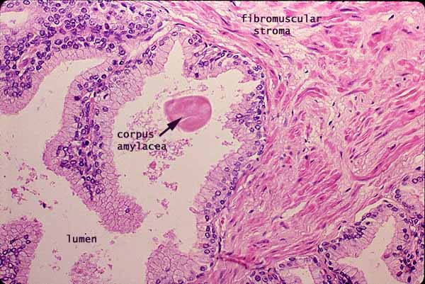
Example 19
Image above: Normal prostate
WebPath example: Chronic prostatitis. [Link opens in new tab.]
In the WebPath image, glandular epithelium appears normal, but the stroma is littered with inflammatory cells.REMINDER: Close tab for WebPath example.
TOP OF PAGE / INDEX OF EXAMPLES / BASIC VOCABULARY
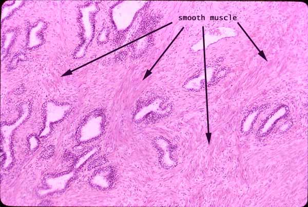
Example 20
Image above: Normal prostate gland. Arrangement of glandular tubules is extremely various in normal prostate, depending on just where the sample is taken; compare with example 21 below.
WebPath example: Adenocarcinoma of prostate. [Link opens in new tab.]
In the WebPath image, the malignant tissue is at left, normal at rightREMINDER: Close tab for WebPath example.
TOP OF PAGE / INDEX OF EXAMPLES / BASIC VOCABULARY
Example 21
Image above: Normal prostate gland
WebPath example: Adenocarcinoma of prostate. [Link opens in new tab.]
Following the arrow link at lower right on the WebPath page will display additional cellular details.REMINDER: Close tab for WebPath example.
TOP OF PAGE / INDEX OF EXAMPLES / BASIC VOCABULARY
Example 22
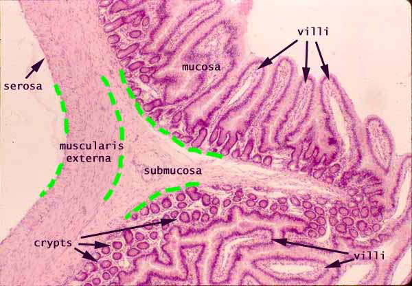
Image above: Small intestine
Example from WikiMedia Commons: Endometriosis in muscularis of intestinal wall. [Link opens in new tab.]
Normal small-intestinal mucosa and submucosa occupy the upper half of the example image. In the lower half, uterine tissue with endometrial glands appears within the muscularis externa.REMINDER: Close tab for pathology example.
TOP OF PAGE / INDEX OF EXAMPLES / BASIC VOCABULARY
Example 23
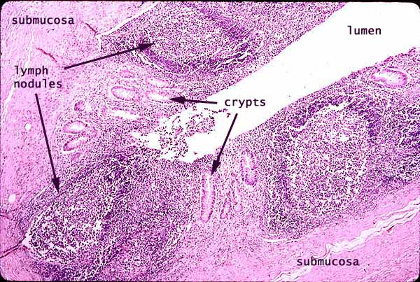
Image above: Normal (post-mortem) appendix
WebPath example: Appendicitis. [Link opens in new tab.]
The WebPath image shows inflammatory infiltrate throughout the wall of the organ, as well as necrosis of the mucosa. In the normal appendix (image above), the abundance of lymphoid tissue might (erroneously) suggest inflammation, but this lymphoid tissue normally includes well-organized lymph nodules.REMINDER: Close tab for WebPath example.
TOP OF PAGE / INDEX OF EXAMPLES / BASIC VOCABULARY
Example 24
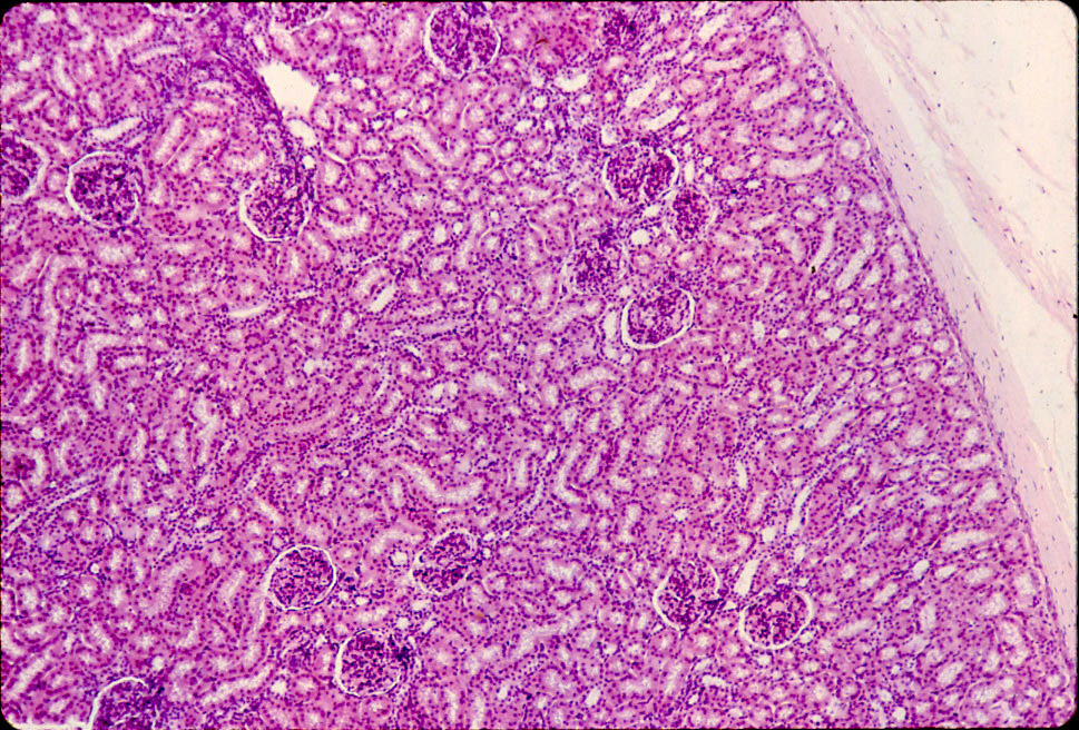
Image above: Normal renal cortex
WebPath example: Acute renal infarct. [Link opens in new tab.]
The WebPath image shows, from left to right, normal, dying, and dead kidney tissue.REMINDER: Close tab for WebPath example.
TOP OF PAGE / INDEX OF EXAMPLES / BASIC VOCABULARY
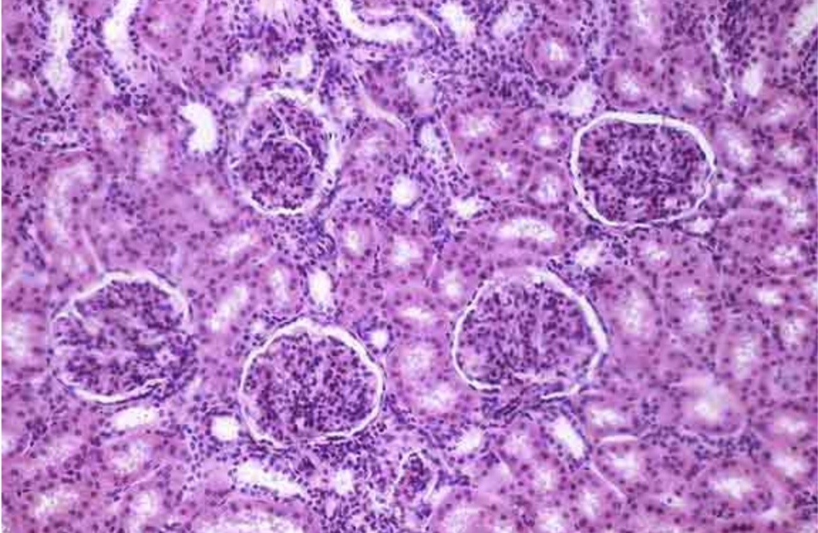
Example 25
Image above: normal renal cortex
WebPath examples, Pyelonephritis [links open in new tabs]:
REMINDER: Close tab for WebPath example.
TOP OF PAGE / INDEX OF EXAMPLES / BASIC VOCABULARY
SPACE FOR ADDITIONAL EXAMPLEExample XX
Image above: caption
WebPath example: XXpathologytopicXX. [Link opens in new tab.]
XXCommentXXREMINDER: Close tab for WebPath example.
TOP OF PAGE / INDEX OF EXAMPLES / BASIC VOCABULARY
External resources for histopathology
- The Internet Pathology Laboratory for Medical Education, hosted by The University of Utah Eccles Health Sciences Library, is a vast resource of materials related to pathology, both gross and histological, including many micrographs of normal and pathological specimens.
- University of Michigan Virtual Slidebox, a large collection of pathology specimens for examination by virtual microscopy.
- Virtual Pathology at the University of Leeds, searchable collection of pathology specimens for examination by virtual microscopy.
- The Pathology Guy: Ed "the Pathguy" Friedlander offers an extensive array of pathology-related resources, including introductory histology.
- For a survey of literature on the application of computational methods for analysis of pathology images, see "Computational Pathology: A Survey Review and The Way Forward," by M.S. Hosseini, et al., at arXiv.
TOP OF PAGE / INDEX OF EXAMPLES / BASIC VOCABULARY
List of WebPath pages linked in examples above.
01. Malignant neoplasm in liver (1).
02. Hepatitis.
03. Cirrhosis.
04. Malignant neoplasm in liver (2).
05. Malignant neoplasm in liver (3).
08. Cervical dysplasia with inflammation.
09. Salivary gland, inflammation with fibrosis.
11. Adenomatous polyp in colon.
12. Pneumonia (1).
13. Pneumonia (2).
14. Lung with metastasized breast carcinoma.
15. Anaphylaxis in nasal mucosa.
16. Squamous metaplasia of larynx.
20. Adenocarcinoma of prostate (low x).
21. Adenocarcinoma of prostate (high x).
22. Endometriosis in intestine.
23. Appendicitis.
24. Renal infarct.
Comments and questions: dgking@siu.edu
SIUC / School
of Medicine / Anatomy / David
King
https://histology.siu.edu/histopath.htm
Last updated: 17 August 2025 / dgk

