

Notes
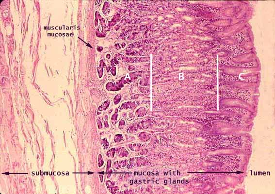
The surface of the stomach is lined by surface mucous cells which comprise a simple columnar epithelium. In the outer (lumenal) region of the mucosa (labelled C in this image), this epithelium is formed into numerous gastric pits. The mucosa beneath the pits is relatively thick and densely packed with simple tubular gasric glands, in the regions labelled A and B above.
In the middle region of the mucosa (B above), each gastric gland is fairly straight and perpendicular to the mucosal surface. In the deeper region of the mucosa (A above), each gland is often more twisted.
At low magnification, such as the image above, the various secretory cells present the appearance of three indistinct bands parallel to the mucosal surface:
- Chief cells predominate in the deep band (A in the figure above);
- Parietal cells predominate in the middle band (B in the figure above); and
- Surface mucous cells line gastric pits in the outer band (C in the figure above).
Mucous neck cells are most common in the necks of the glands (near the line drawn between B and C in the figure above).
Each of these mucosal regions may be examined at higher power:
Related examples:
 |
 |
 |
|
 |
 |
 |
 |
 |
 |
 |
 |
 |
 |
 |
Comments and questions: dgking@siu.edu
SIUC / School
of Medicine / Anatomy / David
King
https://histology.siu.edu/erg/GI100b.htm
Last updated: 27 May 2022 / dgk