
The cervix is a site with special clinical significance, since it is both susceptible to cancer and also relatively accessible for routine diagnostic examination.
The cervix includes the opening (os) from the uterus into the vagina. The tissues associated with this opening are capable of extreme stretching during childbirth.
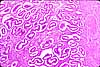
In the vicinity of the os is a transition from the simple columnar, endometrial-type epithelium of the endocervix to the stratified squamous (nonkeratinized) epithelium of the ectocervix, which is continous with that of the vagina. The endocervical mucosa contains many epithelial crevices which confer a glandular appearance.
Before puberty, the epithelial transition occurs near the os. However, enlargement of the uterus and cervix during puberty cause the endocervical mucosa to evert, resulting in a transitional zone of columnar epithelium on the outer surface of the cervix, called the ectropion.
Squamous metaplasia eventually converts this zone into stratified squamous epithelium, but in the process some invaginations of columnar epithelium may lose their connection to the surface. Continuing secretion at such sites may result in the formation of small mucus retention cysts, also known as Nabothian cysts (commemorating Martin Naboth, b. 1675).
The epithelial changes which occur around the cervical os seem to predispose this site to malignant transformation. For examples of cervical dysplasia, see WebPath and WebPath.
The cervical stroma is largely fibrous, with a high proportion of elastic fibers, interwoven with smooth muscle. The stroma is also highly vascularized and richly innervated.
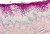
The vagina is lined by non-keratinized stratified squamous epithelium. The epithelial cells accumulate glycogen as they approach the surface.
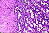
With H&E or trichrome stain, these glycogen-loaded cells appear "empty," with unstained cytoplasmic spaces. With PAS stain the vaginal epithelium stains bright pink/violet.
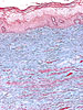
Supporting fibromuscular tissue is highly vascular and richly innervated, with no clear differentiation of mucosa and submucosa (unlike esophagus, which has a superficially similar epithelium). Trichrome stain facilitates observation of smooth muscle.
The vulvar vestibule (between labia majora and labia minora) is lined by stratified squamous epithelium, non-keratized to thinly keratinized. Opening into the vestibule are Bartholin's glands (named for the 17th century Bartholin family of physicians), which secrete lubricating mucus (also see Bartolin's cyst). Located around the opening of the urethra are paraurethral or Skene's glands (named for Scottish gynecologist, Alexander Skene, b. 1837), which are analogous to the male prostate.
 |
 |
|
|
|
|
SIUC / School
of Medicine / Anatomy / David
King
https://histology.siu.edu/erg/cervix.htm
Last updated: 2 February 2023 / dgk