Histology Topics for Lincoln Scholars ProgramWhat is Histology? (an unconventional introduction)
(For a more formal introduction, click here.)
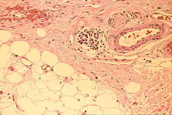
Connective tissues of dermis and hypodermis
Histology is the study of tissues.
The discipline of "histology" takes its name from histo (ιστο), the Greek word for "web." This discipline focuses on the tissue level of anatomical organization.Histology is quite closely related to "microscopic anatomy," and microscopes have indeed become iconic tools for observing tissues. Nevertheless, it is possible to study elaborate microscopic details in the lung, say, or the kidney, without ever appreciating the fundamental tissue organization of each structure's various parts. In contrast, a tissue-level perspective can clarify underlying patterns -- for example, that both lung and kidney share with pancreas a basic pattern of glandular tissue architecture. Or that inflammation can follow the same tissue pathway into many seemingly different organs.
Histology is also closely related to cell biology. But modern cell biology has more in common with biochemistry and molecular biology than with the tissue composition of organs. Cell biology looks inward, into the component parts of individual cells. One can learn a great deal about cells without ever appreciating how cells are arranged into the common set of patterns that underlie the tissue-scale architecture of glands and other organs.
Historical note: Histology emerged historically from recognition that organs consist of several distinct kinds of "stuff." That a tissue-level perspective could offer clinical insight (e.g., by contributing to an understanding of pathological processes such as inflammation) was demonstrated by Xavier Bichat, several decades before establishment of Cell Theory. Contemporary appreciation for histology's value in medicine was nicely captured by George Elliot in her 1871 historical novel Middlemarch:
"That great Frenchman [i.e., Bichat, the "father of histology"] first carried out the conception that living bodies ... must be regarded as consisting of certain primary webs or tissues, out of which the various organs ... are compacted, as the various accommodations of a house are built up in various proportions of wood, iron, stone, brick, zinc, and the rest... And the conception wrought out by Bichat, with his detailed study of the different tissues, acted necessarily on medical questions as the turning of gas-light would act on a dim, oil-lit street, showing new connections and hitherto hidden facts of structure which must be taken into account in considering the symptoms of maladies and the action of medicaments."Bichat described twenty-one "simple tissues." Modern histology has condensed this list into four "basic tissue types": epithelial tissue, connective tissue, muscle tissue, and nervous tissue.
The modern concept of four basic tissue types provides a powerful framework for organizing a great wealth of anatomical and functional detail. Each tissue type has its own characteristic pattern of organization. Yet within each of these basic patterns there are several distinctive varieties:
- Nervous tissue can present as peripheral nerves or peripheral ganglia, or as gray matter and white matter in the central nervous system.
- Muscle comes in three distinct types: smooth, cardiac, and skeletal.
- Epithelial tissue comes in several varieties, each with its own characteristic properties. Yet each of these is distinctively epithelial.
- Connective tissue also comes in several strikingly different forms (including blood and bone as well as "ordinary" connective tissue) that nevertheless share a deep commonality.
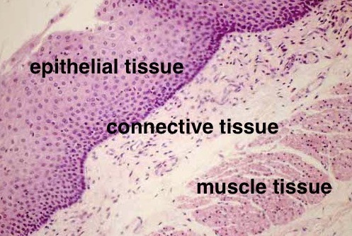
Esophageal mucosa
The basic tissue types can sometimes be readily distinguished from one another, as displayed by tissue layers in the image at right. However, the basic tissue types can also present with a variety of appearances that are not so visually obvious, calling for more subtle interpretation. This is especially true when three-dimensional tissue configurations are sliced and viewed in various planes of section on a microscope slide.
Several varieties of tissue-structures are quite widely distributed throughout the body. Each of the examples in the list below has its own distinctive microscopic appearance, which should eventually become familiar in the context of various organs:
- Ordinary loose connective tissue (also called areolar tissue) occurs around and within practically every organ of the body and forms a readily accessible pathway for inflammation.
- Blood vessels (properly, organs rather than tissues) typically travel within areolar tissue. They are found wherever areolar tissue occurs.
- Adipose connective tissue is also commonly found amidst areolar tissue.
- Columnar epithelium lines many hollow organs.
- Cuboidal epithelium characterizes glands.
- Stratified squamous epithelium forms protective surfaces.
- Peripheral nerves wend their way, accompanied by areolar tissue, into most parts of the body.
- Smooth muscle forms a layer around most hollow organs.
- Skeletal muscle generally occurs in bulk masses of very long and relatively large multi-nucleate fibers. (Cardiac muscle, found only in the heart, is similar but with smaller single-cell fibers.)
♥ The varied appearances of structures in this list comprise a basic "visual vocabulary" for looking at practically any slide.
Additional special varieties of the four basic tissue types occur in a more limited number of locations and can be learned as occasion arises. These include, among others, bone, cartilage, skeletal and cardiac muscle, urothelium (or transitional epithelium), peripheral ganglia, central nervous tissue, lymphoid tissue, and simple squamous epithelium (including mesothelium and endothelium).
For a much longer and more formal introduction to histology, see this page.
Histology Topics for Lincoln Scholars Program
Comments and questions: dgking@siu.edu
SIUC / School
of Medicine / Anatomy / David
King
https://histology.siu.edu/altintro.htm
Last updated: 20 December 2024 / dgk