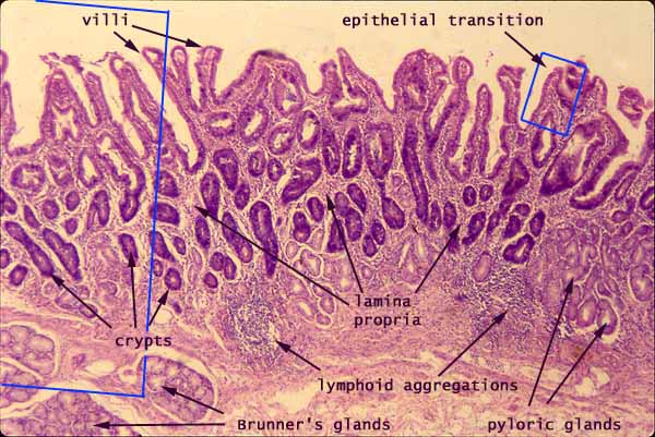

Junction between Stomach and Duodenum, with Pyloric and Brunner's Glands

Notes
Click in any rectangle to magnify.
This slide shows the transition between the stomach (on the far right) and the small intestine (on the left).
Note how mucosal glands on the right give way to submucosal glands on the left. The muscularis mucosa is inconspicuous on this image, but it lies below the pyloric glands and the lymphoid aggregations (on the right) and above the Brunner's glands (on the left).
Click here or in the partial rectangle on left side to see more of the intestine. Click here for more of the stomach.
Click on the small rectangle for a magnified image of the transition between gastric and intestinal epithelium, or...
Click here to see intestinal epithelium, or ...
Click here to see stomach epithelium, or ...
Click here for a very low magnification overview of the stomach-duodenum junction:
Related examples:
 |
 |
 |
 |
 |
 |
 |
 |
 |
 |
 |
 |
 |
 |
 |
Comments and questions: dgking@siu.edu
SIUC / School
of Medicine / Anatomy / David
King
https://histology.siu.edu/erg/GI075b.htm
Last updated: 27 May 2022 / dgk