
Notes
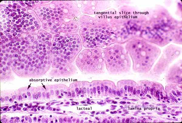
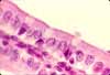
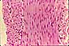
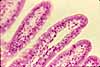
Each villus of the small intestine is lined by simple columnar epithelium composed primarily of absorptive cells (enterocytes), with scattered goblet cells.
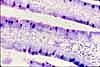
A villus appears in standard longitudinal section in the lower third of this image, displaying, its simple columnar epithelium, its core of lamina propria, and its lacteal.
In the upper two-thirds of the image, the plane of section shaves through the epithelium without going deeper into lamina propria. Such a section provides a different perspective on the arrangement of the epithelial cells.
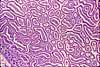
The arrangement of small, round nuclei demonstrates how the columnar absorptive epithelial cells are closely packed together in a hexagonal array.
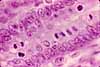
The empty "bubbles" (white arrows in the inset to right) are mucous droplets of goblet cells. Such a plane of section provides a good sense of the relative numbers of goblet cells vs. absorptive cells, and also suggests how bulging goblets may disturb the neat arrangement of absorptive cells.
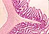
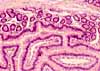
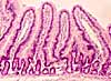
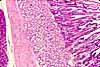
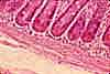
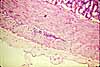
More small intestine examples:
 |
 |
 |
 |
 |
 |
 |
 |
< |
 |
 |
 |
Comments and questions: dgking@siu.edu
SIUC / School
of Medicine / Anatomy / David
King
https://histology.siu.edu/erg/GI145b.htm
Last updated: 27 May 2022 / dgk