 The
thick mucosa of the stomach is characterized
by closely packed tubular glands beneath a surface of simple columnar epithelium. Lamina propria is highly vascular, with capillaries embracing each gland.
 The
surface is indented into numerous short gastric
pits which open freely to the lumen. The entire surface
consists uniformly of surface mucous cells, which
protect the stomach from self-digestion.
Failure of the surface mucous cells to protect the stomach
wall can lead to an ulcer.
 Gastric
glands comprise the bulk of the mucosa beneath the pits. Although
lamina propria separates the individual glands, most of the mucosal volume
is occupied by secretory cells, primarily parietal
cells and chief cells.
The submucosa of the stomach is relatively
unspecialized.

The muscularis externa of the stomach
is thicker than elsewhere along the tract. The smooth muscle fibers
of the muscularis may form an oblique layer in addition to the inner circular
and outer longitudinal" layers that charaterize the rest of the tract.
Multiple layers are often difficult to distinguish microscopically,
because in any given section only one of the layers (at most) will be cut
in cross section while the others will be more or less oblique. An especially thick
region forms the pyloric sphincter
As elsewhere along the tract, the serosa
of the stomach is normally unremarkable.
GASTRIC PITS and GASTRIC GLANDS
 With
their tubular shape and mucous-secretory surface, gastric
pits have a distinctly glandular appearance. However, since one
single cell type, the surface mucous cell,
is continuous over the entire stomach surface, the pits are usually regarded
not as glands but just as shallow indentations of the surface epithelium.
Several gastric glands may open into the bottom of each gastric pit.
 Beneath
the gastric pits, the mucosa of the stomach is filled with closely packed
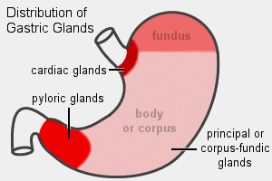
tubular glands, which differ
by region (i.e., cardiac, fundic, pyloric). Histologically, most
of the stomach wall contains gastric glands (or fundic glands). These
consist primarily of parietal cells and
chief cells. The fundic glands also
contain mucous neck cells and stem
cells.
 Gastric
parietal cells (oxyntic cells) secrete
acid, by pumping chloride and hydrogen ions. These cells are unique
not only in function but also in microscopic appearance. Parietal
cells may be found at any level in the fundic glands, but they are most
plentiful in the middle region of the mucosa.
 Gastric
chief cells secrete the digestive enzymes
and have typical serous-secretory appearance.
Chief cells may be found at any level in the fundic glands, but they
are most plentiful in the deeper mucosa, toward the muscularis
mucosae.
 Mucous
neck cells are inconspicuous cells with a typical
mucous-secretory
appearance. These cells are most common in the upper region of the
fundic glands. Their specific function remains unclear.
Stem cells,
located near the point where gastric glands open into gastric pits, are
responsible for replenishing the gland cells and also the surface
mucous cells that protect the stomach surface. These cells are
difficult to notice and even more difficult to identify in routine histological
preparations.
 Microscopic
appearance of fundic glands. In routine sections, the arrangement
of secretory cells into tubular glands is often not readily apparent. The
cells may appear to be all jumbled together, or associated into cords, for
several reasons:
- The glands are not quite straight and not quite parallel
to one another.
- The cells which comprise the glands vary in size, shape
and appearance.
- There is often little or no free space in the glandular
lumens.
 Since
tubular organization often cannot be clearly seen, the orientation of the
secretory cells is also often obscure. Nevertheless, the predominant
secretory cells (parietal cells and chief
cells) may be recognized as epithelial by their typical round, euchromatic
nuclei. The glandular architecture usually appears most neatly where
chief cells predominate, deep in the mucosa near the mucularis mucosae.
 Individual
glands are separated from one another by a thin stroma of lamina
propria. The lamina propria
may be recognized by the densely heterochromatic, irregular or flattened nuclei
of its fibroblasts and endothelial cells. This lamina
propria contains the capillaries which nourish the glands and the stomach
surface and which also supply the materials needed for secretion.
 The
junction between esophagus and stomach is marked by
a sharp transition from the stratified squamous epithelium of the esophageal
mucosa to the simple columnar epithelium
of the gastric mucosa.
The mucosa of the stomach is divided, somewhat
arbitrarily, into three regions: the cardiac stomach, the fundic
stomach, and the pyloric stomach.
|

Image from Blue
Histology,
Copyright Lutz Slomianka 1998-2004
|
These anatomical regions differ histologically primarily
in the length and composition of the mucosal glands.
Cardiac glands are relatively short, relative to the
fundic and pyloric glands.
Fundic glands are described above.
 Pyloric
glands are especially extensive. These glands are functionally and
structurally similar to Brunner's glands
of the duodenum. Both are tubular mucous
glands which secrete alkaline mucus. These mucous glands remain within the mucosa
in the stomach but expand across the muscularis
mucosae to fill the submucosa of the
duodenum.
Both cardiac and pyloric glands are predominantly
mucous (i.e., few parietal or chief cells). This makes functional
sense, since mucus can help protect the adjacent regions of the tract from
damage by stomach acid and digestive enzymes. Surface
mucous cells protect the stomach lining, but the esophagus
and duodenum lack any such protection.
The
pyloric stomach is also characterized by a considerable thickening of the
muscularis externa, forming the pyloric
sphincter.
 The junction between stomach and duodenum is marked not only by the pyloric sphincter
but also by an abrupt change in mucosal tissue. The gastric epithelium of surface
mucous cells (lining gastric pits) ends at the beginning of intestinal epithelium of absorptive
cells (covering villi). And the pyloric mucosal glands expand into the submucosa of the
duodenum.

 The
thick mucosa of the stomach is characterized
by closely packed tubular glands beneath a surface of simple columnar epithelium. Lamina propria is highly vascular, with capillaries embracing each gland.
The surface is indented into numerous short gastric pits which open freely to the lumen. The entire surface consists uniformly of surface mucous cells, which protect the stomach from self-digestion.
Gastric glands comprise the bulk of the mucosa beneath the pits. Although lamina propria separates the individual glands, most of the mucosal volume is occupied by secretory cells, primarily parietal cells and chief cells.


