

Notes
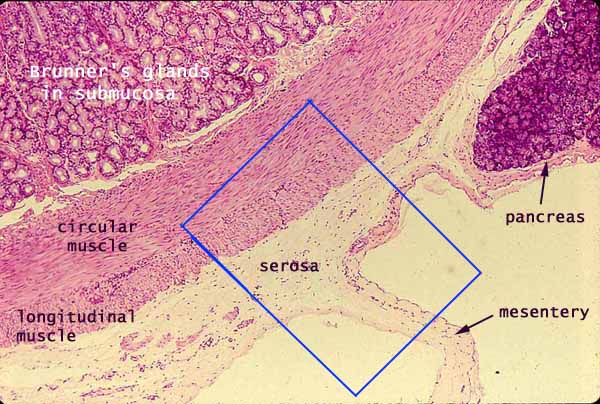
The serosa consists of connective tissue with a delicate covering of mesothelium (simple squamous epithelium derived from mesoderm). The serosa is continuous with the mesentery. Click here or within the rectangle to magnify the region of the serosa and mesentery.
The muscularis externa of the small intestine consists of smooth muscle fibers arranged into an inner circular layer and an outer longitudinal layer. The appearance of smooth muscle varies dramatically, depending on the orientation of the fibers with respect to the plane of section.
In this section, the circular muscle fibers are cut longitudinally, while the longitudinal muscle fibers are cut in cross section (transversely). (More on appearance of smooth muscle.)
Between these two muscle layers is a network of unmyelinated nerve fibers and parasympathetic ganglia called Auerbach's plexus (or myenteric plexus).
Note the submucosa at upper right in this micrograph appears to be packed with mucous glands. Which segment of the small intestine has this submucosal appearance?
Related examples:
 |
 |
 |
 |
 |
 |
 |
 |
 |
 |
 |
 |
 |
 |
 |
 |
 |
 |
 |
 |
 |
 |
 |
 |
Comments and questions: dgking@siu.edu
SIUC / School
of Medicine / Anatomy / David
King
https://histology.siu.edu/erg/GI001b.htm
Last updated: 27 May 2022 / dgk